 Movie
Movie Controller


+ Open data

- Basic information
| Entry | Database: PDB / ID: 3zxn | ||||||
|---|---|---|---|---|---|---|---|
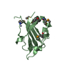
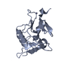
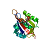
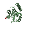
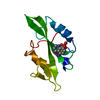
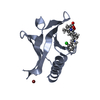
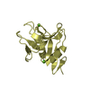
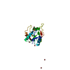
| Title | Moorella thermoacetica RsbS S58E | ||||||
 Components Components | ANTI-SIGMA-FACTOR ANTAGONIST (STAS) DOMAIN PROTEIN | ||||||
 Keywords Keywords |  TRANSCRIPTION / GENE REGULATION TRANSCRIPTION / GENE REGULATION | ||||||
| Function / homology |  Function and homology information Function and homology informationSTAS domain / STAS domain / Transcription Regulator spoIIAA / STAS domain / STAS domain profile. / STAS domain / STAS domain superfamily / 2-Layer Sandwich / Alpha Beta Similarity search - Domain/homology | ||||||
| Biological species |   MOORELLA THERMOACETICA (bacteria) MOORELLA THERMOACETICA (bacteria) | ||||||
| Method | X-RAY DIFFRACTION / SYNCHROTRON / MOLECULAR REPLACEMENT / Resolution: 1.9 Å | ||||||
 Authors Authors | Quin, M.B. / Berrisford, J.M. / Newman, J.A. / Basle, A. / Lewis, R.J. / Marles-Wright, J. | ||||||
 Citation Citation | Journal: Structure / Year: 2012 Title: The Bacterial Stressosome: A Modular System that Has Been Adapted to Control Secondary Messenger Signaling. Authors: Quin, M.B. / Berrisford, J.M. / Newman, J.A. / Basle, A. / Lewis, R.J. / Marles-Wright, J. | ||||||
| History |
|
- Structure visualization
| Structure viewer | Molecule: MolmilJmol/JSmol |
|---|
- Downloads & links
-Download
| PDBx/mmCIF format | 3zxn.cif.gz | 61.2 KB | Display | PDBx/mmCIF format |
|---|---|---|---|---|
| PDB format | pdb3zxn.ent.gz | 45.3 KB | Display | PDB format |
| PDBx/mmJSON format | 3zxn.json.gz | Tree view | PDBx/mmJSON format | |
| Others |  Other downloads Other downloads |
-Validation report
| Arichive directory | https://data.pdbj.org/pub/pdb/validation_reports/zx/3zxnftp://data.pdbj.org/pub/pdb/validation_reports/zx/3zxn | HTTPS FTP |
|---|
-Related structure data
| Related structure data | 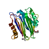 3zt9C 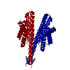 3ztaC 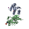 3ztbC  2vy9S C: citing same article ( S: Starting model for refinement |
|---|---|
| Similar structure data |


-Links
 PDBj
PDBj

- Assembly
| Deposited unit | 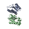
| ||||||||
|---|---|---|---|---|---|---|---|---|---|
| 1 | 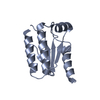
| ||||||||
| 2 | 
| ||||||||
| Unit cell |
|
-Components
| #1: Protein | Mass: 13623.000 Da / Num. of mol.: 2 / Mutation: YES Source method: isolated from a genetically manipulated source Source: (gene. exp.) MOORELLA THERMOACETICA (bacteria) / Description: GERMAN COLLECTION OF MICROORGANISMS (DSM) / Production host: ESCHERICHIA COLI (E. coli) / Strain (production host): BL21(DE3) / References: UniProt: Q2RIF5#2: Chemical | Thiocyanate  Mass: 58.082 Da / Num. of mol.: 2 / Source method: obtained synthetically / Formula: CNS Mass: 58.082 Da / Num. of mol.: 2 / Source method: obtained synthetically / Formula: CNS#3: Water | ChemComp-HOH / | Water Mass: 18.015 Da / Num. of mol.: 169 / Source method: isolated from a natural source / Formula: H2O Mass: 18.015 Da / Num. of mol.: 169 / Source method: isolated from a natural source / Formula: H2OCompound details | ENGINEERED | |
|---|
-Experimental details
-Experiment
| Experiment | Method: X-RAY DIFFRACTION / Number of used crystals: 1 |
|---|
- Sample preparation
| Crystal | Density Matthews: 2.66 Å3/Da / Density % sol: 53.81 % / Description: NONE |
|---|---|
| Crystal grow | pH: 7 Details: 0.1 M HEPES PH7, 8% (W/V) PEG 8000, 0.1 M SODIUM THIOCYANATE |
-Data collection
| Diffraction | Mean temperature: 100 K |
|---|---|
| Diffraction source | Source: SYNCHROTRON / Site: Diamond  / Beamline: I02 / Wavelength: 0.979 / Beamline: I02 / Wavelength: 0.979 |
| Detector | Type: ADSC CCD / Detector: CCD / Date: May 30, 2011 |
| Radiation | Protocol: SINGLE WAVELENGTH / Monochromatic (M) / Laue (L): M / Scattering type: x-ray |
| Radiation wavelength | Wavelength: 0.979 Å / Relative weight: 1 |
| Reflection | Resolution: 1.9→49.94 Å / Num. obs: 21867 / % possible obs: 99.4 % / Observed criterion σ(I): -3 / Redundancy: 3.6 % / Biso Wilson estimate: 30.263 Å2 / Rmerge(I) obs: 0.06 / Net I/σ(I): 12.2 |
| Reflection shell | Resolution: 1.9→2 Å / Redundancy: 3.7 % / Rmerge(I) obs: 0.37 / Mean I/σ(I) obs: 2.8 / % possible all: 99.9 |
- Processing
| Software |
| ||||||||||||||||||||||||||||||||||||||||||||||||||||||||||||||||||||||||||||||||||||||||||||||||||||||||||||||||
|---|---|---|---|---|---|---|---|---|---|---|---|---|---|---|---|---|---|---|---|---|---|---|---|---|---|---|---|---|---|---|---|---|---|---|---|---|---|---|---|---|---|---|---|---|---|---|---|---|---|---|---|---|---|---|---|---|---|---|---|---|---|---|---|---|---|---|---|---|---|---|---|---|---|---|---|---|---|---|---|---|---|---|---|---|---|---|---|---|---|---|---|---|---|---|---|---|---|---|---|---|---|---|---|---|---|---|---|---|---|---|---|---|---|
| Refinement | Method to determine structure: MOLECULAR REPLACEMENT Starting model: PDB ENTRY 2VY9 Resolution: 1.9→29.144 Å / SU ML: 0.25 / σ(F): 1.25 / Phase error: 24.27 / Stereochemistry target values: ML Details: RESIDUES 1-2, 23 AND 121 TO 123 ARE DISORDERED IN CHAIN A. RESIDUES 1-4 AND RESIDUES 119 TO 123 ARE DISORDERED IN CHAIN B.
| ||||||||||||||||||||||||||||||||||||||||||||||||||||||||||||||||||||||||||||||||||||||||||||||||||||||||||||||||
| Solvent computation | Shrinkage radii: 0.83 Å / VDW probe radii: 1.1 Å / Solvent model: FLAT BULK SOLVENT MODEL / Bsol: 73.257 Å2 / ksol: 0.385 e/Å3 | ||||||||||||||||||||||||||||||||||||||||||||||||||||||||||||||||||||||||||||||||||||||||||||||||||||||||||||||||
| Displacement parameters | Biso mean: 38.582 Å2
| ||||||||||||||||||||||||||||||||||||||||||||||||||||||||||||||||||||||||||||||||||||||||||||||||||||||||||||||||
| Refinement step | Cycle: LAST / Resolution: 1.9→29.144 Å
| ||||||||||||||||||||||||||||||||||||||||||||||||||||||||||||||||||||||||||||||||||||||||||||||||||||||||||||||||
| Refine LS restraints |
| ||||||||||||||||||||||||||||||||||||||||||||||||||||||||||||||||||||||||||||||||||||||||||||||||||||||||||||||||
| LS refinement shell |
|
