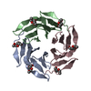
 Movie
Movie Controller


+ Open data

- Basic information
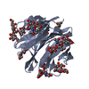
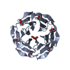
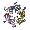
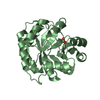
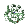
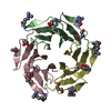
| Entry | Database: PDB / ID: 3zw0 | ||||||
|---|---|---|---|---|---|---|---|
| Title | Structure of BambL lectin from Burkholderia ambifaria | ||||||
 Components Components | (BAMBL LECTIN) x 2 | ||||||
 Keywords Keywords | SUGAR BINDING PROTEIN /  CYSTIC FIBROSIS / B-PROPELLER / HUMAN HISTO-BLOOD GROUP CYSTIC FIBROSIS / B-PROPELLER / HUMAN HISTO-BLOOD GROUP | ||||||
| Function / homology | Lipocalin - #190 / Fucose-specific lectin / Fungal fucose-specific lectin / Lipocalin / Beta Barrel / Mainly Beta / alpha-L-fucopyranose / Fucose-binding lectin protein Function and homology information Function and homology information | ||||||
| Biological species |  BURKHOLDERIA AMBIFARIA (bacteria) BURKHOLDERIA AMBIFARIA (bacteria) | ||||||
| Method | X-RAY DIFFRACTION / SYNCHROTRON / MOLECULAR REPLACEMENT / Resolution: 1.6 Å | ||||||
 Authors Authors | Audfray, A. / Claudinon, J. / Abounit, S. / Ruvoen-Clouet, N. / Larson, G. / Wimmerova, M. / LePendu, J. / Romer, W. / Varrot, A. / Imberty, A. | ||||||
 Citation Citation | Journal: J.Biol.Chem. / Year: 2012 Title: Fucose-Binding Lectin from Opportunistic Pathogen Burkholderia Ambifaria Binds to Both Plant and Human Oligosaccharidic Epitopes. Authors: Audfray, A. / Claudinon, J. / Abounit, S. / Ruvoen-Clouet, N. / Larson, G. / Smith, D.F. / Wimmerova, M. / Le Pendu, J. / Romer, W. / Varrot, A. / Imberty, A. | ||||||
| History |
| ||||||
| Remark 700 | SHEET DETERMINATION METHOD: AUTHOR PROVIDED. |
- Structure visualization
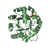
| Structure viewer | Molecule: MolmilJmol/JSmol |
|---|
- Downloads & links
-Download
| PDBx/mmCIF format | 3zw0.cif.gz | 72.6 KB | Display | PDBx/mmCIF format |
|---|---|---|---|---|
| PDB format | pdb3zw0.ent.gz | 53.1 KB | Display | PDB format |
| PDBx/mmJSON format | 3zw0.json.gz | Tree view | PDBx/mmJSON format | |
| Others |  Other downloads Other downloads |
-Validation report
| Arichive directory | https://data.pdbj.org/pub/pdb/validation_reports/zw/3zw0ftp://data.pdbj.org/pub/pdb/validation_reports/zw/3zw0 | HTTPS FTP |
|---|
-Related structure data
| Related structure data |  3zw2C  3zweC  3zzvC 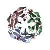 2bt9S C: citing same article ( S: Starting model for refinement |
|---|---|
| Similar structure data |


-Links
 PDBj
PDBj
- Assembly
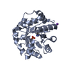
| Deposited unit | 
| ||||||||||||
|---|---|---|---|---|---|---|---|---|---|---|---|---|---|
| 1 |
| ||||||||||||
| Unit cell |
| ||||||||||||
| Noncrystallographic symmetry (NCS) | NCS oper:
|
-Components
| #1: Protein | Mass: 9387.166 Da / Num. of mol.: 2 Source method: isolated from a genetically manipulated source Source: (gene. exp.) BURKHOLDERIA AMBIFARIA (bacteria) / Strain: AMMD / Production host: ESCHERICHIA COLI (E. coli) / Strain (production host): BL21(DE3) / References: UniProt: Q0B4G1#2: Protein | | Mass: 9403.166 Da / Num. of mol.: 1 Source method: isolated from a genetically manipulated source Source: (gene. exp.) BURKHOLDERIA AMBIFARIA (bacteria) / Strain: AMMD / Production host: ESCHERICHIA COLI (E. coli) / Strain (production host): BL21(DE3) / References: UniProt: Q0B4G1#3: Sugar | ChemComp-FUC / | Fucose  Type: L-saccharide, alpha linking / Mass: 164.156 Da / Num. of mol.: 1 Type: L-saccharide, alpha linking / Mass: 164.156 Da / Num. of mol.: 1Source method: isolated from a genetically manipulated source Formula: C6H12O5 #4: Water | ChemComp-HOH / | Water Mass: 18.015 Da / Num. of mol.: 399 / Source method: isolated from a natural source / Formula: H2O Mass: 18.015 Da / Num. of mol.: 399 / Source method: isolated from a natural source / Formula: H2O |
|---|
-Experimental details
-Experiment
| Experiment | Method: X-RAY DIFFRACTION / Number of used crystals: 1 |
|---|
- Sample preparation
| Crystal | Density Matthews: 1.78 Å3/Da / Density % sol: 31.11 % / Description: NONE |
|---|---|
| Crystal grow | pH: 7.5 / Details: pH 7.5 |
-Data collection
| Diffraction | Mean temperature: 100 K |
|---|---|
| Diffraction source | Source: SYNCHROTRON / Site: ESRF  / Beamline: BM30A / Wavelength: 0.9334 / Beamline: BM30A / Wavelength: 0.9334 |
| Detector | Type: ADSC CCD / Detector: CCD / Date: Jul 16, 2010 |
| Radiation | Protocol: SINGLE WAVELENGTH / Monochromatic (M) / Laue (L): M / Scattering type: x-ray |
| Radiation wavelength | Wavelength: 0.9334 Å / Relative weight: 1 |
| Reflection | Resolution: 1.6→33.24 Å / Num. obs: 26647 / % possible obs: 93.7 % / Observed criterion σ(I): 3 / Redundancy: 3.7 % / Biso Wilson estimate: 10.2 Å2 / Rmerge(I) obs: 0.03 / Net I/σ(I): 26.6 |
| Reflection shell | Resolution: 1.6→1.69 Å / Redundancy: 2.6 % / Rmerge(I) obs: 0.06 / Mean I/σ(I) obs: 13.4 / % possible all: 70.5 |
- Processing
| Software |
| ||||||||||||||||||||||||||||||||||||||||||||||||||||||||||||||||||||||||||||||||||||||||||||||||||||||||||||||||||||||||||||||||||||||||||||||||||||||||||||||||||||||||||||||||||||||
|---|---|---|---|---|---|---|---|---|---|---|---|---|---|---|---|---|---|---|---|---|---|---|---|---|---|---|---|---|---|---|---|---|---|---|---|---|---|---|---|---|---|---|---|---|---|---|---|---|---|---|---|---|---|---|---|---|---|---|---|---|---|---|---|---|---|---|---|---|---|---|---|---|---|---|---|---|---|---|---|---|---|---|---|---|---|---|---|---|---|---|---|---|---|---|---|---|---|---|---|---|---|---|---|---|---|---|---|---|---|---|---|---|---|---|---|---|---|---|---|---|---|---|---|---|---|---|---|---|---|---|---|---|---|---|---|---|---|---|---|---|---|---|---|---|---|---|---|---|---|---|---|---|---|---|---|---|---|---|---|---|---|---|---|---|---|---|---|---|---|---|---|---|---|---|---|---|---|---|---|---|---|---|---|
| Refinement | Method to determine structure: MOLECULAR REPLACEMENT Starting model: PDB ENTRY 2BT9 Resolution: 1.6→49.16 Å / Cor.coef. Fo:Fc: 0.962 / Cor.coef. Fo:Fc free: 0.935 / SU B: 1.537 / SU ML: 0.055 / Cross valid method: THROUGHOUT / ESU R: 0.095 / ESU R Free: 0.098 / Stereochemistry target values: MAXIMUM LIKELIHOOD Details: HYDROGENS HAVE BEEN ADDED IN THE RIDING POSITIONS. U VALUES REFINED INDIVIDUALLY.
| ||||||||||||||||||||||||||||||||||||||||||||||||||||||||||||||||||||||||||||||||||||||||||||||||||||||||||||||||||||||||||||||||||||||||||||||||||||||||||||||||||||||||||||||||||||||
| Solvent computation | Ion probe radii: 0.8 Å / Shrinkage radii: 0.8 Å / VDW probe radii: 1.4 Å / Solvent model: MASK | ||||||||||||||||||||||||||||||||||||||||||||||||||||||||||||||||||||||||||||||||||||||||||||||||||||||||||||||||||||||||||||||||||||||||||||||||||||||||||||||||||||||||||||||||||||||
| Displacement parameters | Biso mean: 7.177 Å2
| ||||||||||||||||||||||||||||||||||||||||||||||||||||||||||||||||||||||||||||||||||||||||||||||||||||||||||||||||||||||||||||||||||||||||||||||||||||||||||||||||||||||||||||||||||||||
| Refinement step | Cycle: LAST / Resolution: 1.6→49.16 Å
| ||||||||||||||||||||||||||||||||||||||||||||||||||||||||||||||||||||||||||||||||||||||||||||||||||||||||||||||||||||||||||||||||||||||||||||||||||||||||||||||||||||||||||||||||||||||
| Refine LS restraints |
|