[English] 日本語
Yorodumi
- PDB-3zuc: Structure of CBM3b of major scaffoldin subunit ScaA from Acetivib... -

+
Open data


ID or keywords:

Loading...

-
Basic information

Entry
Database: PDB / ID: 3zuc
Title
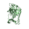
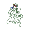
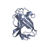
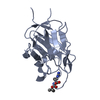
Structure of CBM3b of major scaffoldin subunit ScaA from Acetivibrio cellulolyticus determined from the crystals grown in the presence of Nickel
ComponentsCELLULOSOMAL SCAFFOLDIN
KeywordsCRYSTALLINE CELLULOSE-BINDING PROTEIN / SUGAR BINDING PROTEIN / CELLULOSOME
Function / homology
Function and homology information


cellulose binding / cellulase / cellulase activity / cellulose catabolic process / extracellular region / metal ion binding
Similarity search - Function
Endoglucanase-like / CarboxypepD_reg-like domain / Glycosyl hydrolases family 9, Asp/Glu active sites / Glycosyl hydrolases family 9 (GH9) active site signature 3. / Cellulosome anchoring protein, cohesin domain / Cohesin domain / Carboxypeptidase-like, regulatory domain superfamily / Cellulose binding domain / Cellulose binding domain / Carbohydrate-binding module 3 ...Endoglucanase-like / CarboxypepD_reg-like domain / Glycosyl hydrolases family 9, Asp/Glu active sites / Glycosyl hydrolases family 9 (GH9) active site signature 3. / Cellulosome anchoring protein, cohesin domain / Cohesin domain / Carboxypeptidase-like, regulatory domain superfamily / Cellulose binding domain / Cellulose binding domain / Carbohydrate-binding module 3 / Carbohydrate-binding module 3 superfamily / CBM3 (carbohydrate binding type-3) domain profile. / Glycoside hydrolase family 9 / Glycosyl hydrolase family 9 / Dockerin domain / Dockerin domain profile. / Dockerin domain superfamily / CBM2/CBM3, carbohydrate-binding domain superfamily / Six-hairpin glycosidase-like superfamily / Six-hairpin glycosidase superfamily / EF-Hand 1, calcium-binding site / EF-hand calcium-binding domain. / Immunoglobulin-like / Sandwich / Mainly Beta
Similarity search - Domain/homology
NICKEL (II) ION / Endoglucanase
Similarity search - Component
Biological speciesACETIVIBRIO CELLULOLYTICUS (bacteria)
MethodX-RAY DIFFRACTION / SYNCHROTRON / MOLECULAR REPLACEMENT / Resolution: 1.001 Å
AuthorsYaniv, O. / Halfon, Y. / Lamed, R. / Frolow, F.
CitationJournal: Acta Crystallogr.,Sect.F / Year: 2012
Title: Structure of Cbm3B of the Major Scaffoldin Subunit Scaa from Acetivibrio Cellulolyticus
Authors: Yaniv, O. / Halfon, Y. / Shimon, L.J.W. / Bayer, E.A. / Lamed, R. / Frolow, F.
History
DepositionJul 18, 2011Deposition site: PDBE / Processing site: PDBE
Revision 1.0Jan 11, 2012Provider: repository / Type: Initial release
Revision 1.1Apr 11, 2012Group: Other
Revision 1.2Mar 29, 2017Group: Structure summary
Revision 2.0Dec 20, 2023Group: Atomic model / Data collection ...Atomic model / Data collection / Database references / Derived calculations / Other / Refinement description
Category: atom_site / chem_comp_atom ...atom_site / chem_comp_atom / chem_comp_bond / database_2 / pdbx_database_status / pdbx_initial_refinement_model / pdbx_struct_conn_angle / pdbx_struct_special_symmetry / struct_conn / struct_site
Item: _atom_site.B_iso_or_equiv / _atom_site.Cartn_x ..._atom_site.B_iso_or_equiv / _atom_site.Cartn_x / _atom_site.Cartn_y / _atom_site.Cartn_z / _database_2.pdbx_DOI / _database_2.pdbx_database_accession / _pdbx_database_status.status_code_sf / _pdbx_struct_conn_angle.ptnr1_auth_comp_id / _pdbx_struct_conn_angle.ptnr1_auth_seq_id / _pdbx_struct_conn_angle.ptnr1_label_asym_id / _pdbx_struct_conn_angle.ptnr1_label_atom_id / _pdbx_struct_conn_angle.ptnr1_label_comp_id / _pdbx_struct_conn_angle.ptnr1_label_seq_id / _pdbx_struct_conn_angle.ptnr3_auth_comp_id / _pdbx_struct_conn_angle.ptnr3_auth_seq_id / _pdbx_struct_conn_angle.ptnr3_label_asym_id / _pdbx_struct_conn_angle.ptnr3_label_atom_id / _pdbx_struct_conn_angle.ptnr3_label_comp_id / _pdbx_struct_conn_angle.ptnr3_label_seq_id / _pdbx_struct_conn_angle.value / _struct_conn.pdbx_dist_value / _struct_conn.ptnr1_auth_comp_id / _struct_conn.ptnr1_auth_seq_id / _struct_conn.ptnr1_label_asym_id / _struct_conn.ptnr1_label_atom_id / _struct_conn.ptnr1_label_comp_id / _struct_conn.ptnr1_label_seq_id / _struct_conn.ptnr2_auth_comp_id / _struct_conn.ptnr2_auth_seq_id / _struct_conn.ptnr2_label_asym_id / _struct_conn.ptnr2_label_atom_id / _struct_conn.ptnr2_label_comp_id / _struct_conn.ptnr2_label_seq_id / _struct_site.pdbx_auth_asym_id / _struct_site.pdbx_auth_comp_id / _struct_site.pdbx_auth_seq_id

-
Structure visualization

Structure viewerMolecule:
MolmilJmol/JSmol

Downloads & links

-
Assembly

Deposited unit
A: CELLULOSOMAL SCAFFOLDIN
hetero molecules


Theoretical massNumber of molelcules
Total (without water)17,1875
Polymers16,7881
Non-polymers3994
Water5,603311
1


  • Idetical with deposited unit
  • defined by author&software
TypeNameSymmetry operationNumber
identity operation1_555x,y,z1
MethodPISA
Unit cell
Length a, b, c (Å)52.697, 52.697, 194.159
Angle α, β, γ (deg.)90.00, 90.00, 120.00
Int Tables number178
Space group name H-MP6122
Components on special symmetry positions
IDModelComponents
11A-2101-

HOH

21A-2311-

HOH

-
Components

-
Protein , 1 types, 1 molecules A

#1: Protein CELLULOSOMAL SCAFFOLDIN / FAMILY 3B CARBOHYDRATE BINDING MODULE


Mass: 16788.248 Da / Num. of mol.: 1 / Fragment: RESIDUES 973-1121
Source method: isolated from a genetically manipulated source
Source: (gene. exp.) ACETIVIBRIO CELLULOLYTICUS (bacteria) / Production host: ESCHERICHIA COLI (E. coli) / Strain (production host): BL21 / Variant (production host): RIL / References: UniProt: Q9RPL0

-
Non-polymers , 5 types, 315 molecules

#2: Chemical ChemComp-CA / CALCIUM ION


Mass: 40.078 Da / Num. of mol.: 1 / Source method: obtained synthetically / Formula: Ca
#3: Chemical ChemComp-EDO / 1,2-ETHANEDIOL / ETHYLENE GLYCOL / Ethylene glycol


Mass: 62.068 Da / Num. of mol.: 1 / Source method: obtained synthetically / Formula: C2H6O2
#4: Chemical ChemComp-NI / NICKEL (II) ION / Nickel


Mass: 58.693 Da / Num. of mol.: 1 / Source method: obtained synthetically / Formula: Ni
#5: Chemical ChemComp-1PE / PENTAETHYLENE GLYCOL / PEG400 / Polyethylene glycol


Mass: 238.278 Da / Num. of mol.: 1 / Source method: obtained synthetically / Formula: C10H22O6 / Comment: precipitant*YM
#6: Water ChemComp-HOH / water / Water


Mass: 18.015 Da / Num. of mol.: 311 / Source method: isolated from a natural source / Formula: H2O

-
Details

Nonpolymer detailsNICKEL (II) ION (NI): CRYSTALS WERE GROWN IN THE PRESENCE OF NICKEL IONS

-
Experimental details

-
Experiment

ExperimentMethod: X-RAY DIFFRACTION / Number of used crystals: 1

-
Sample preparation

CrystalDensity Matthews: 2.24 Å3/Da / Density % sol: 45.2 % / Description: NONE
Crystal growpH: 7.5
Details: 1.9 M AMMONIUM SULFATE,0.1 M HEPES PH 7.5 0.5% (W/V) POLYETHYLENE GLYCOL 400 0.2M NICKEL CHLORIDE

-
Data collection

DiffractionMean temperature: 100 K
Diffraction sourceSource: SYNCHROTRON / Site: ESRF / Beamline: ID29 / Wavelength: 0.97625
DetectorType: DECTRIS PILATUS 6M / Detector: PIXEL / Date: Jun 25, 2011 / Details: MIRRORS
RadiationMonochromator: SI(311) MONOCHROMATOR / Protocol: SINGLE WAVELENGTH / Monochromatic (M) / Laue (L): M / Scattering type: x-ray
Radiation wavelengthWavelength: 0.97625 Å / Relative weight: 1
ReflectionResolution: 1→50 Å / Num. obs: 87445 / % possible obs: 98.4 % / Observed criterion σ(I): 0 / Redundancy: 7.6 % / Biso Wilson estimate: 8.24 Å2 / Rmerge(I) obs: 0.05 / Net I/σ(I): 33.2
Reflection shellResolution: 1→1.02 Å / Redundancy: 5.8 % / Rmerge(I) obs: 0.57 / Mean I/σ(I) obs: 1.6 / % possible all: 82.2

-
Processing

Software
NameVersionClassification
PHENIX(PHENIX.REFINE)refinement
MOSFLMdata reduction
SCALEPACKdata scaling
MOLREPphasing
RefinementMethod to determine structure: MOLECULAR REPLACEMENT
Starting model: PDB ENTRY 3ZQW
Resolution: 1.001→44.426 Å / SU ML: 0.21 / σ(F): 1.35 / Phase error: 10.62 / Stereochemistry target values: ML
Details: 159400 REFLECTIONS USED IN REFINEMENT USING SEPARATED FRIEDEL PAIRS
RfactorNum. reflection% reflection
Rfree0.136 3719 2.3 %
Rwork0.1249 --
obs0.1251 159400 98.08 %
Solvent computationShrinkage radii: 0.6 Å / VDW probe radii: 0.45 Å / Solvent model: FLAT BULK SOLVENT MODEL / Bsol: 75.879 Å2 / ksol: 0.498 e/Å3
Displacement parametersBiso mean: 12.8 Å2
Baniso -1Baniso -2Baniso -3
1--1.41 Å20 Å20 Å2
2---1.41 Å20 Å2
3---2.82 Å2
Refinement stepCycle: LAST / Resolution: 1.001→44.426 Å
ProteinNucleic acidLigandSolventTotal
Num. atoms1187 0 22 311 1520
Refine LS restraints
Refine-IDTypeDev idealNumber
X-RAY DIFFRACTIONf_bond_d0.0111369
X-RAY DIFFRACTIONf_angle_d1.4021877
X-RAY DIFFRACTIONf_dihedral_angle_d14.715479
X-RAY DIFFRACTIONf_chiral_restr0.101195
X-RAY DIFFRACTIONf_plane_restr0.009249
LS refinement shell
Resolution (Å)Rfactor RfreeNum. reflection RfreeRfactor RworkNum. reflection RworkRefine-ID% reflection obs (%)
1.0011-1.01370.35911120.34674611X-RAY DIFFRACTION77
1.0137-1.02710.2731210.29815144X-RAY DIFFRACTION88
1.0271-1.04110.28181340.25695454X-RAY DIFFRACTION93
1.0411-1.0560.24391350.22065677X-RAY DIFFRACTION96
1.056-1.07180.18121410.18785816X-RAY DIFFRACTION99
1.0718-1.08850.16981380.14835796X-RAY DIFFRACTION99
1.0885-1.10640.14261370.13065806X-RAY DIFFRACTION99
1.1064-1.12550.14711360.12115852X-RAY DIFFRACTION99
1.1255-1.14590.13571350.10885866X-RAY DIFFRACTION99
1.1459-1.1680.12041420.10665839X-RAY DIFFRACTION100
1.168-1.19180.13071440.10165844X-RAY DIFFRACTION100
1.1918-1.21770.11991440.10255854X-RAY DIFFRACTION100
1.2177-1.24610.11721400.10045850X-RAY DIFFRACTION100
1.2461-1.27720.10541430.1015857X-RAY DIFFRACTION100
1.2772-1.31180.12651410.10015886X-RAY DIFFRACTION100
1.3118-1.35040.11641440.09885904X-RAY DIFFRACTION100
1.3504-1.3940.10441380.09755850X-RAY DIFFRACTION100
1.394-1.44380.10431370.09565848X-RAY DIFFRACTION100
1.4438-1.50160.1111390.09285942X-RAY DIFFRACTION100
1.5016-1.56990.11881390.09385823X-RAY DIFFRACTION100
1.5699-1.65270.12121380.09295911X-RAY DIFFRACTION100
1.6527-1.75630.10531430.15883X-RAY DIFFRACTION100
1.7563-1.89190.11591400.10315850X-RAY DIFFRACTION100
1.8919-2.08220.1311360.10495890X-RAY DIFFRACTION100
2.0822-2.38350.11031350.11745871X-RAY DIFFRACTION100
2.3835-3.00290.16211460.13755861X-RAY DIFFRACTION100
3.0029-44.470.14931410.1595896X-RAY DIFFRACTION100

+
About Yorodumi

-
News

-
Feb 9, 2022. New format data for meta-information of EMDB entries

New format data for meta-information of EMDB entries

  • Version 3 of the EMDB header file is now the official format.
  • The previous official version 1.9 will be removed from the archive.

Related info.:EMDB header

External links:wwPDB to switch to version 3 of the EMDB data model

-
Aug 12, 2020. Covid-19 info

Covid-19 info

URL: https://pdbj.org/emnavi/covid19.php

New page: Covid-19 featured information page in EM Navigator.

Related info.:Covid-19 info / Mar 5, 2020. Novel coronavirus structure data

+
Mar 5, 2020. Novel coronavirus structure data

Novel coronavirus structure data

Related info.:Yorodumi Speices / Aug 12, 2020. Covid-19 info

External links:COVID-19 featured content - PDBj / Molecule of the Month (242):Coronavirus Proteases

+
Jan 31, 2019. EMDB accession codes are about to change! (news from PDBe EMDB page)

EMDB accession codes are about to change! (news from PDBe EMDB page)

  • The allocation of 4 digits for EMDB accession codes will soon come to an end. Whilst these codes will remain in use, new EMDB accession codes will include an additional digit and will expand incrementally as the available range of codes is exhausted. The current 4-digit format prefixed with “EMD-” (i.e. EMD-XXXX) will advance to a 5-digit format (i.e. EMD-XXXXX), and so on. It is currently estimated that the 4-digit codes will be depleted around Spring 2019, at which point the 5-digit format will come into force.
  • The EM Navigator/Yorodumi systems omit the EMD- prefix.

Related info.:Q: What is EMD? / ID/Accession-code notation in Yorodumi/EM Navigator

External links:EMDB Accession Codes are Changing Soon! / Contact to PDBj

+
Jul 12, 2017. Major update of PDB

Major update of PDB

  • wwPDB released updated PDB data conforming to the new PDBx/mmCIF dictionary.
  • This is a major update changing the version number from 4 to 5, and with Remediation, in which all the entries are updated.
  • In this update, many items about electron microscopy experimental information are reorganized (e.g. em_software).
  • Now, EM Navigator and Yorodumi are based on the updated data.

External links:wwPDB Remediation / Enriched Model Files Conforming to OneDep Data Standards Now Available in the PDB FTP Archive

-
Yorodumi

Thousand views of thousand structures

  • Yorodumi is a browser for structure data from EMDB, PDB, SASBDB, etc.
  • This page is also the successor to EM Navigator detail page, and also detail information page/front-end page for Omokage search.
  • The word "yorodu" (or yorozu) is an old Japanese word meaning "ten thousand". "mi" (miru) is to see.

Related info.:EMDB / PDB / SASBDB / Comparison of 3 databanks / Yorodumi Search / Aug 31, 2016. New EM Navigator & Yorodumi / Yorodumi Papers / Jmol/JSmol / Function and homology information / Changes in new EM Navigator and Yorodumi

Read more