[English] 日本語
Yorodumi
- PDB-3zpp: Structure of the Streptococcus pneumoniae surface protein and adh... -

+
Open data


ID or keywords:

Loading...

-
Basic information

Entry
Database: PDB / ID: 3zpp
TitleStructure of the Streptococcus pneumoniae surface protein and adhesin PfbA
ComponentsCELL WALL SURFACE ANCHOR FAMILY PROTEIN
Keywords
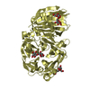
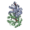
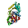
CELL ADHESION / PFBA / SURFACE ADHESIN / SP1833 / MSCRAMM
Function / homology
Function and homology information


cell wall / extracellular region / membrane / metal ion binding
Similarity search - Function
Right handed beta helix domain / Right handed beta helix region / Pectate lyase superfamily protein / Pectate lyase superfamily protein / Parallel beta-helix repeat / Parallel beta-helix repeats / Single-stranded right-handed beta-helix, Pectin lyase-like / Pectate Lyase C-like / YSIRK type signal peptide / Pectin lyase fold ...Right handed beta helix domain / Right handed beta helix region / Pectate lyase superfamily protein / Pectate lyase superfamily protein / Parallel beta-helix repeat / Parallel beta-helix repeats / Single-stranded right-handed beta-helix, Pectin lyase-like / Pectate Lyase C-like / YSIRK type signal peptide / Pectin lyase fold / YSIRK Gram-positive signal peptide / Pectin lyase fold/virulence factor / 3 Solenoid / Gram-positive cocci surface proteins LPxTG motif profile. / LPXTG cell wall anchor domain / Mainly Beta
Similarity search - Domain/homology
Cell wall surface anchor family protein / Cell wall surface anchor family protein
Similarity search - Component
Biological speciesSTREPTOCOCCUS PNEUMONIAE (bacteria)
MethodX-RAY DIFFRACTION / SYNCHROTRON / MOLECULAR REPLACEMENT / Resolution: 2.28 Å
AuthorsL Suits, M.D. / Boraston, A.B.
CitationJournal: Plos One / Year: 2013
Title: Structure of the Streptococcus Pneumoniae Surface Protein and Adhesin Pfba.
Authors: Suits, M.D. / Boraston, A.B.
History
DepositionFeb 28, 2013Deposition site: PDBE / Processing site: PDBE
Revision 1.0May 29, 2013Provider: repository / Type: Initial release
Revision 1.1Aug 14, 2013Group: Database references
Revision 1.2May 8, 2024Group: Data collection / Database references ...Data collection / Database references / Derived calculations / Other
Category: chem_comp_atom / chem_comp_bond ...chem_comp_atom / chem_comp_bond / database_2 / pdbx_database_status / pdbx_struct_conn_angle / struct_conn / struct_site
Item: _database_2.pdbx_DOI / _database_2.pdbx_database_accession ..._database_2.pdbx_DOI / _database_2.pdbx_database_accession / _pdbx_database_status.status_code_sf / _pdbx_struct_conn_angle.ptnr1_auth_comp_id / _pdbx_struct_conn_angle.ptnr1_auth_seq_id / _pdbx_struct_conn_angle.ptnr1_label_asym_id / _pdbx_struct_conn_angle.ptnr1_label_comp_id / _pdbx_struct_conn_angle.ptnr1_label_seq_id / _pdbx_struct_conn_angle.ptnr3_auth_comp_id / _pdbx_struct_conn_angle.ptnr3_auth_seq_id / _pdbx_struct_conn_angle.ptnr3_label_asym_id / _pdbx_struct_conn_angle.ptnr3_label_comp_id / _pdbx_struct_conn_angle.ptnr3_label_seq_id / _pdbx_struct_conn_angle.value / _struct_conn.pdbx_dist_value / _struct_conn.ptnr1_auth_comp_id / _struct_conn.ptnr1_auth_seq_id / _struct_conn.ptnr1_label_asym_id / _struct_conn.ptnr1_label_atom_id / _struct_conn.ptnr1_label_comp_id / _struct_conn.ptnr1_label_seq_id / _struct_conn.ptnr2_auth_comp_id / _struct_conn.ptnr2_auth_seq_id / _struct_conn.ptnr2_label_asym_id / _struct_conn.ptnr2_label_atom_id / _struct_conn.ptnr2_label_comp_id / _struct_conn.ptnr2_label_seq_id / _struct_site.pdbx_auth_asym_id / _struct_site.pdbx_auth_comp_id / _struct_site.pdbx_auth_seq_id

-
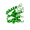
Structure visualization

Structure viewerMolecule:
MolmilJmol/JSmol

Downloads & links

-
Assembly

Deposited unit
A: CELL WALL SURFACE ANCHOR FAMILY PROTEIN
hetero molecules


Theoretical massNumber of molelcules
Total (without water)48,4442
Polymers48,4041
Non-polymers401
Water1,06359
1


  • Idetical with deposited unit
  • defined by author&software
TypeNameSymmetry operationNumber
identity operation1_555x,y,z1
MethodPISA
Unit cell
Length a, b, c (Å)62.840, 140.650, 128.640
Angle α, β, γ (deg.)90.00, 90.00, 90.00
Int Tables number20
Space group name H-MC2221

-
Components

#1: Protein CELL WALL SURFACE ANCHOR FAMILY PROTEIN / SP_1833


Mass: 48404.105 Da / Num. of mol.: 1 / Fragment: CORE-DOMAIN, RESIDUES 139-560
Source method: isolated from a genetically manipulated source
Source: (gene. exp.) STREPTOCOCCUS PNEUMONIAE (bacteria) / Strain: TIGR4 / Plasmid: PET28A / Production host: ESCHERICHIA COLI (E. coli) / Strain (production host): BL21(DE3) / References: UniProt: Q97P11, UniProt: A0A0H2URN9*PLUS
#2: Chemical ChemComp-CA / CALCIUM ION


Mass: 40.078 Da / Num. of mol.: 1 / Source method: obtained synthetically / Formula: Ca
#3: Water ChemComp-HOH / water / Water


Mass: 18.015 Da / Num. of mol.: 59 / Source method: isolated from a natural source / Formula: H2O
Sequence detailsTHIS IS A TRUNCATED FORM OF THE PROTEIN SPANNING RESIDUES 139-560.

-
Experimental details

-
Experiment

ExperimentMethod: X-RAY DIFFRACTION / Number of used crystals: 1

-
Sample preparation

CrystalDensity Matthews: 2.94 Å3/Da / Density % sol: 58 %
Description: STRUCTURE WAS SOLVED VIA MR FROM A SAD SELENOMETHIONINE DERIVATIVE.
Crystal growpH: 5.5
Details: 12% (W/V) 10K POLYETHYLENE GLYCOL, 80 MM MG DIFORMATE (C2H2MGO4) AND BIS-TRIS-HCL (PH 5.5)

-
Data collection

DiffractionMean temperature: 100 K
Diffraction sourceSource: SYNCHROTRON / Site: CLSI / Beamline: 08ID-1 / Wavelength: 0.97949
DetectorType: MARMOSAIC 225 mm CCD / Detector: CCD / Date: Oct 15, 2012 / Details: VERTICAL COLLIMATING MIRROR
RadiationMonochromator: DOUBLE CRYSTAL MONOCHROMATOR (DCM) / Protocol: SINGLE WAVELENGTH / Monochromatic (M) / Laue (L): M / Scattering type: x-ray
Radiation wavelengthWavelength: 0.97949 Å / Relative weight: 1
ReflectionResolution: 2.28→38 Å / Num. obs: 26446 / % possible obs: 100 % / Redundancy: 5.5 % / Rmerge(I) obs: 0.13 / Net I/σ(I): 6.7
Reflection shellResolution: 2.28→2.4 Å / Redundancy: 5.5 % / Rmerge(I) obs: 0.5 / Mean I/σ(I) obs: 2.4 / % possible all: 100

-
Processing

Software
NameVersionClassification
REFMAC5.7.0029refinement
XDSdata reduction
SCALAdata scaling
PHASERphasing
RefinementMethod to determine structure: MOLECULAR REPLACEMENT / Resolution: 2.28→57.44 Å / Cor.coef. Fo:Fc: 0.956 / Cor.coef. Fo:Fc free: 0.934 / SU B: 7.253 / SU ML: 0.171 / Cross valid method: THROUGHOUT / ESU R: 0.256 / ESU R Free: 0.215 / Stereochemistry target values: MAXIMUM LIKELIHOOD
Details: HYDROGENS HAVE BEEN ADDED IN THE RIDING POSITIONS. HYDROGENS HAVE BEEN USED IF PRESENT IN THE INPUT. U VALUES REFINED INDIVIDUALLY N-TERMINAL IMAC AFFINITY TAG IS PRESENT IN THE CONSTRUCT ...Details: HYDROGENS HAVE BEEN ADDED IN THE RIDING POSITIONS. HYDROGENS HAVE BEEN USED IF PRESENT IN THE INPUT. U VALUES REFINED INDIVIDUALLY N-TERMINAL IMAC AFFINITY TAG IS PRESENT IN THE CONSTRUCT BUT NOT MODELED INTO THE COORDINATES DUE TO THE ABSENCE OF CLEAR ELECTRON DENSITY FOR THIS REGION.
RfactorNum. reflection% reflectionSelection details
Rfree0.2551 1340 5.1 %RANDOM
Rwork0.20804 ---
obs0.21045 25097 99.97 %-
Solvent computationIon probe radii: 0.8 Å / Shrinkage radii: 0.8 Å / VDW probe radii: 1.2 Å / Solvent model: MASK
Displacement parametersBiso mean: 42.156 Å2
Baniso -1Baniso -2Baniso -3
1-5.26 Å20 Å20 Å2
2--1.04 Å20 Å2
3----6.29 Å2
Refinement stepCycle: LAST / Resolution: 2.28→57.44 Å
ProteinNucleic acidLigandSolventTotal
Num. atoms3236 0 1 59 3296
Refine LS restraints
Refine-IDTypeDev idealDev ideal targetNumber
X-RAY DIFFRACTIONr_bond_refined_d0.0130.0193311
X-RAY DIFFRACTIONr_bond_other_d
X-RAY DIFFRACTIONr_angle_refined_deg1.6661.9484481
X-RAY DIFFRACTIONr_angle_other_deg
X-RAY DIFFRACTIONr_dihedral_angle_1_deg8.795428
X-RAY DIFFRACTIONr_dihedral_angle_2_deg43.60326.346156
X-RAY DIFFRACTIONr_dihedral_angle_3_deg18.50515587
X-RAY DIFFRACTIONr_dihedral_angle_4_deg13.727158
X-RAY DIFFRACTIONr_chiral_restr0.1190.2508
X-RAY DIFFRACTIONr_gen_planes_refined0.010.022501
X-RAY DIFFRACTIONr_gen_planes_other
X-RAY DIFFRACTIONr_nbd_refined
X-RAY DIFFRACTIONr_nbd_other
X-RAY DIFFRACTIONr_nbtor_refined
X-RAY DIFFRACTIONr_nbtor_other
X-RAY DIFFRACTIONr_xyhbond_nbd_refined
X-RAY DIFFRACTIONr_xyhbond_nbd_other
X-RAY DIFFRACTIONr_metal_ion_refined
X-RAY DIFFRACTIONr_metal_ion_other
X-RAY DIFFRACTIONr_symmetry_vdw_refined
X-RAY DIFFRACTIONr_symmetry_vdw_other
X-RAY DIFFRACTIONr_symmetry_hbond_refined
X-RAY DIFFRACTIONr_symmetry_hbond_other
X-RAY DIFFRACTIONr_symmetry_metal_ion_refined
X-RAY DIFFRACTIONr_symmetry_metal_ion_other
X-RAY DIFFRACTIONr_mcbond_it
X-RAY DIFFRACTIONr_mcbond_other
X-RAY DIFFRACTIONr_mcangle_it
X-RAY DIFFRACTIONr_mcangle_other
X-RAY DIFFRACTIONr_scbond_it
X-RAY DIFFRACTIONr_scbond_other
X-RAY DIFFRACTIONr_scangle_it
X-RAY DIFFRACTIONr_scangle_other
X-RAY DIFFRACTIONr_long_range_B_refined
X-RAY DIFFRACTIONr_long_range_B_other
X-RAY DIFFRACTIONr_rigid_bond_restr
X-RAY DIFFRACTIONr_sphericity_free
X-RAY DIFFRACTIONr_sphericity_bonded
LS refinement shellResolution: 2.28→2.339 Å / Total num. of bins used: 20
RfactorNum. reflection% reflection
Rfree0.349 85 -
Rwork0.306 1859 -
obs--100 %

+
About Yorodumi

-
News

-
Feb 9, 2022. New format data for meta-information of EMDB entries

New format data for meta-information of EMDB entries

  • Version 3 of the EMDB header file is now the official format.
  • The previous official version 1.9 will be removed from the archive.

Related info.:EMDB header

External links:wwPDB to switch to version 3 of the EMDB data model

-
Aug 12, 2020. Covid-19 info

Covid-19 info

URL: https://pdbj.org/emnavi/covid19.php

New page: Covid-19 featured information page in EM Navigator.

Related info.:Covid-19 info / Mar 5, 2020. Novel coronavirus structure data

+
Mar 5, 2020. Novel coronavirus structure data

Novel coronavirus structure data

Related info.:Yorodumi Speices / Aug 12, 2020. Covid-19 info

External links:COVID-19 featured content - PDBj / Molecule of the Month (242):Coronavirus Proteases

+
Jan 31, 2019. EMDB accession codes are about to change! (news from PDBe EMDB page)

EMDB accession codes are about to change! (news from PDBe EMDB page)

  • The allocation of 4 digits for EMDB accession codes will soon come to an end. Whilst these codes will remain in use, new EMDB accession codes will include an additional digit and will expand incrementally as the available range of codes is exhausted. The current 4-digit format prefixed with “EMD-” (i.e. EMD-XXXX) will advance to a 5-digit format (i.e. EMD-XXXXX), and so on. It is currently estimated that the 4-digit codes will be depleted around Spring 2019, at which point the 5-digit format will come into force.
  • The EM Navigator/Yorodumi systems omit the EMD- prefix.

Related info.:Q: What is EMD? / ID/Accession-code notation in Yorodumi/EM Navigator

External links:EMDB Accession Codes are Changing Soon! / Contact to PDBj

+
Jul 12, 2017. Major update of PDB

Major update of PDB

  • wwPDB released updated PDB data conforming to the new PDBx/mmCIF dictionary.
  • This is a major update changing the version number from 4 to 5, and with Remediation, in which all the entries are updated.
  • In this update, many items about electron microscopy experimental information are reorganized (e.g. em_software).
  • Now, EM Navigator and Yorodumi are based on the updated data.

External links:wwPDB Remediation / Enriched Model Files Conforming to OneDep Data Standards Now Available in the PDB FTP Archive

-
Yorodumi

Thousand views of thousand structures

  • Yorodumi is a browser for structure data from EMDB, PDB, SASBDB, etc.
  • This page is also the successor to EM Navigator detail page, and also detail information page/front-end page for Omokage search.
  • The word "yorodu" (or yorozu) is an old Japanese word meaning "ten thousand". "mi" (miru) is to see.

Related info.:EMDB / PDB / SASBDB / Comparison of 3 databanks / Yorodumi Search / Aug 31, 2016. New EM Navigator & Yorodumi / Yorodumi Papers / Jmol/JSmol / Function and homology information / Changes in new EM Navigator and Yorodumi

Read more