 Movie
Movie Controller

[English] 日本語
 Yorodumi
- PDB-3zgx: Crystal structure of the kleisin-N SMC interface in prokaryotic c... -
+ Open data

- Basic information
| Entry | Database: PDB / ID: 3zgx | ||||||
|---|---|---|---|---|---|---|---|
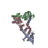
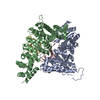
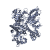
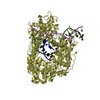
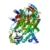
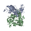
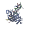
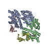
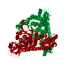
| Title | Crystal structure of the kleisin-N SMC interface in prokaryotic condensin | ||||||
 Components Components |
| ||||||
 Keywords Keywords |  CELL CYCLE CELL CYCLE | ||||||
| Function / homology |  Function and homology information Function and homology informationchromosome condensation / sister chromatid cohesion / chromosome segregation / chromosome / DNA replication / cell division / ATP hydrolysis activity / DNA binding / ATP binding / identical protein binding / cytoplasmSimilarity search - Function | ||||||
| Biological species |  BACILLUS SUBTILIS (bacteria) BACILLUS SUBTILIS (bacteria) | ||||||
| Method | X-RAY DIFFRACTION / SYNCHROTRON / SAD / Resolution: 3.4 Å | ||||||
 Authors Authors | Burmann, F. / Shin, H. / Basquin, J. / Soh, Y. / Gimenez, V. / Kim, Y. / Oh, B. / Gruber, S. | ||||||
 Citation Citation | Journal: Nat.Struct.Mol.Biol. / Year: 2013 Title: An Asymmetric Smc-Kleisin Bridge in Prokaryotic Condensin. Authors: Burmann, F. / Shin, H. / Basquin, J. / Soh, Y. / Gimenez-Oya, V. / Kim, Y. / Oh, B. / Gruber, S. | ||||||
| History |
|
- Structure visualization
| Structure viewer | Molecule: MolmilJmol/JSmol |
|---|
- Downloads & links
-Download
| PDBx/mmCIF format | 3zgx.cif.gz | 172.4 KB | Display | PDBx/mmCIF format |
|---|---|---|---|---|
| PDB format | pdb3zgx.ent.gz | 135.3 KB | Display | PDB format |
| PDBx/mmJSON format | 3zgx.json.gz | Tree view | PDBx/mmJSON format | |
| Others |  Other downloads Other downloads |
-Validation report
| Arichive directory | https://data.pdbj.org/pub/pdb/validation_reports/zg/3zgxftp://data.pdbj.org/pub/pdb/validation_reports/zg/3zgx | HTTPS FTP |
|---|
-Related structure data


-Links
 PDBj
PDBj- Assembly
| Deposited unit | 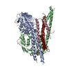
| |||||||||||||||||||||||||||||||||
|---|---|---|---|---|---|---|---|---|---|---|---|---|---|---|---|---|---|---|---|---|---|---|---|---|---|---|---|---|---|---|---|---|---|---|
| 1 |
| |||||||||||||||||||||||||||||||||
| Unit cell |
| |||||||||||||||||||||||||||||||||
| Noncrystallographic symmetry (NCS) | NCS domain:
NCS domain segments:
NCS ensembles :
|
-Components
| #1: Protein | Mass: 48166.711 Da / Num. of mol.: 2 / Fragment: SMC HEAD DOMAIN, RESIDUES 1-219,983-1186 Source method: isolated from a genetically manipulated source Source: (gene. exp.) BACILLUS SUBTILIS (bacteria) / Strain: 1A700 / Plasmid: PET28 POLYCISTRONIC / Production host: ESCHERICHIA COLI (E. coli) / Strain (production host): BL21 / Variant (production host): GOLD / References: UniProt: P51834#2: Protein | Mass: 11182.849 Da / Num. of mol.: 2 / Fragment: N TERMINAL DOMAIN OF SCPA, RESIDUES 1-86 Source method: isolated from a genetically manipulated source Source: (gene. exp.) BACILLUS SUBTILIS (bacteria) / Strain: 1A700 / Plasmid: PET 28 POLYCISTRONIC / Production host: ESCHERICHIA COLI (E. coli) / Strain (production host): BL21 / Variant (production host): GOLD / References: UniProt: P35154Sequence details | C TERMINAL HEXAHISTID | |
|---|
-Experimental details
-Experiment
| Experiment | Method: X-RAY DIFFRACTION / Number of used crystals: 1 |
|---|
- Sample preparation
| Crystal | Density Matthews: 2.54 Å3/Da / Density % sol: 51.5 % / Description: NONE |
|---|---|
| Crystal grow | pH: 6.5 Details: 8 % ISOPROPANOL, 20 MM MAGNESIUM CHLORIDE AND 50 MM MES PH 6.5 |
-Data collection
| Diffraction | Mean temperature: 100 K |
|---|---|
| Diffraction source | Source: SYNCHROTRON / Site: SLS  / Beamline: X10SA / Wavelength: 0.9793 / Beamline: X10SA / Wavelength: 0.9793 |
| Detector | Type: DECTRIS PILATUS 6M / Detector: PIXEL / Date: Dec 10, 2011 |
| Radiation | Protocol: SINGLE WAVELENGTH / Monochromatic (M) / Laue (L): M / Scattering type: x-ray |
| Radiation wavelength | Wavelength: 0.9793 Å / Relative weight: 1 |
| Reflection twin | Operator: h,-k,-l / Fraction: 0.49 |
| Reflection | Resolution: 3.4→75.97 Å / Num. obs: 16156 / % possible obs: 99.9 % / Observed criterion σ(I): 3.3 / Redundancy: 9.5 % / Rmerge(I) obs: 0.09 / Net I/σ(I): 18.8 |
| Reflection shell | Resolution: 3.4→3.58 Å / Redundancy: 9.1 % / Rmerge(I) obs: 0.58 / Mean I/σ(I) obs: 3.3 / % possible all: 99.2 |
- Processing
| Software |
| ||||||||||||||||||||||||||||||||||||||||||||||||||||||||||||||||||||||||||||||||||||
|---|---|---|---|---|---|---|---|---|---|---|---|---|---|---|---|---|---|---|---|---|---|---|---|---|---|---|---|---|---|---|---|---|---|---|---|---|---|---|---|---|---|---|---|---|---|---|---|---|---|---|---|---|---|---|---|---|---|---|---|---|---|---|---|---|---|---|---|---|---|---|---|---|---|---|---|---|---|---|---|---|---|---|---|---|---|
| Refinement | Method to determine structure: SAD Starting model: NONE Resolution: 3.4→75.968 Å / σ(F): 1.99 / Phase error: 33.28 / Stereochemistry target values: TWIN_LSQ_F
| ||||||||||||||||||||||||||||||||||||||||||||||||||||||||||||||||||||||||||||||||||||
| Solvent computation | Shrinkage radii: 0.9 Å / VDW probe radii: 1.11 Å / Solvent model: FLAT BULK SOLVENT MODEL | ||||||||||||||||||||||||||||||||||||||||||||||||||||||||||||||||||||||||||||||||||||
| Refinement step | Cycle: LAST / Resolution: 3.4→75.968 Å
| ||||||||||||||||||||||||||||||||||||||||||||||||||||||||||||||||||||||||||||||||||||
| Refine LS restraints |
| ||||||||||||||||||||||||||||||||||||||||||||||||||||||||||||||||||||||||||||||||||||
| Refine LS restraints NCS |
| ||||||||||||||||||||||||||||||||||||||||||||||||||||||||||||||||||||||||||||||||||||
| LS refinement shell |
|
