 Movie
Movie Controller

[English] 日本語
 Yorodumi
- PDB-3zgb: Greater efficiency of photosynthetic carbon fixation due to singl... -
+ Open data

- Basic information
| Entry | Database: PDB / ID: 3zgb | ||||||
|---|---|---|---|---|---|---|---|
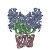
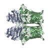
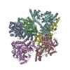
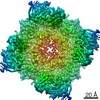
| Title | Greater efficiency of photosynthetic carbon fixation due to single amino acid substitution | ||||||
 Components Components | PHOSPHOENOLPYRUVATE CARBOXYLASE | ||||||
 Keywords Keywords | LYASE | ||||||
| Function / homology |  Function and homology informationphosphoenolpyruvate carboxylase / phosphoenolpyruvate carboxylase activity / carbon fixation / photosynthesis / tricarboxylic acid cycle / cytoplasm Function and homology informationphosphoenolpyruvate carboxylase / phosphoenolpyruvate carboxylase activity / carbon fixation / photosynthesis / tricarboxylic acid cycle / cytoplasmSimilarity search - Function | ||||||
| Biological species |  Flaveria pringlei (plant) Flaveria pringlei (plant) | ||||||
| Method | X-RAY DIFFRACTION / SYNCHROTRON / MOLECULAR REPLACEMENT / Resolution: 2.71 Å | ||||||
 Authors Authors | Paulus, J.K. / Schlieper, D. / Groth, G. | ||||||
 Citation Citation | Journal: Nat.Commun. / Year: 2013 Title: Greater Efficiency of Photosynthetic Carbon Fixation due to Single Amino Acid Substitution Authors: Paulus, J.K. / Schlieper, D. / Groth, G. | ||||||
| History |
| ||||||
| Remark 700 | SHEET DETERMINATION METHOD: DSSP THE SHEETS PRESENTED AS "AA" IN EACH CHAIN ON SHEET RECORDS BELOW ... SHEET DETERMINATION METHOD: DSSP THE SHEETS PRESENTED AS "AA" IN EACH CHAIN ON SHEET RECORDS BELOW IS ACTUALLY AN 8-STRANDED BARREL THIS IS REPRESENTED BY A 9-STRANDED SHEET IN WHICH THE FIRST AND LAST STRANDS ARE IDENTICAL. THE SHEETS PRESENTED AS "BA" IN EACH CHAIN ON SHEET RECORDS BELOW IS ACTUALLY AN 8-STRANDED BARREL THIS IS REPRESENTED BY A 9-STRANDED SHEET IN WHICH THE FIRST AND LAST STRANDS ARE IDENTICAL. |
- Structure visualization
| Structure viewer | Molecule: MolmilJmol/JSmol |
|---|
- Downloads & links
-Download
| PDBx/mmCIF format | 3zgb.cif.gz | 730.8 KB | Display | PDBx/mmCIF format |
|---|---|---|---|---|
| PDB format | pdb3zgb.ent.gz | 608.6 KB | Display | PDB format |
| PDBx/mmJSON format | 3zgb.json.gz | Tree view | PDBx/mmJSON format | |
| Others |  Other downloads Other downloads |
-Validation report
| Arichive directory | https://data.pdbj.org/pub/pdb/validation_reports/zg/3zgbftp://data.pdbj.org/pub/pdb/validation_reports/zg/3zgb | HTTPS FTP |
|---|
-Related structure data
| Related structure data |  3zgeC  3zbeS C: citing same article ( S: Starting model for refinement |
|---|---|
| Similar structure data |


-Links
 PDBj
PDBj

- Assembly
| Deposited unit | 
| ||||||||
|---|---|---|---|---|---|---|---|---|---|
| 1 | 
| ||||||||
| Unit cell |
|
-Components
| #1: Protein | / PEPC / PEPCASE / PPCA Mass: 111472.367 Da / Num. of mol.: 2 Source method: isolated from a genetically manipulated source Details: THE GENE NAME OF THE PROTEIN IS PPCA / Source: (gene. exp.) Flaveria pringlei (plant) / Plasmid: PETEV16B / Production host:  Escherichia coli BL21(DE3) (bacteria) / Variant (production host): GOLD Escherichia coli BL21(DE3) (bacteria) / Variant (production host): GOLDReferences: UniProt: Q01647, phosphoenolpyruvate carboxylase#2: Chemical | Aspartic acid  Type: L-peptide linking / Mass: 133.103 Da / Num. of mol.: 2 / Source method: obtained synthetically / Formula: C4H7NO4 Type: L-peptide linking / Mass: 133.103 Da / Num. of mol.: 2 / Source method: obtained synthetically / Formula: C4H7NO4#3: Chemical | Ethylene glycol  Mass: 62.068 Da / Num. of mol.: 2 / Source method: obtained synthetically / Formula: C2H6O2 Mass: 62.068 Da / Num. of mol.: 2 / Source method: obtained synthetically / Formula: C2H6O2#4: Chemical | ChemComp-SO4 / Sulfate  Mass: 96.063 Da / Num. of mol.: 4 / Source method: obtained synthetically / Formula: SO4 Mass: 96.063 Da / Num. of mol.: 4 / Source method: obtained synthetically / Formula: SO4#5: Water | ChemComp-HOH / | Water Mass: 18.015 Da / Num. of mol.: 56 / Source method: isolated from a natural source / Formula: H2O Mass: 18.015 Da / Num. of mol.: 56 / Source method: isolated from a natural source / Formula: H2OSequence details | THE CRYSTALLIZED SEQUENCE ACTUALLY MAPS TO NCBI ACCESSION CODE Z48966 WITHOUT ANY CONFLICTS OR GAPS. ...THE CRYSTALLIZ | |
|---|
-Experimental details
-Experiment
| Experiment | Method: X-RAY DIFFRACTION / Number of used crystals: 1 |
|---|
- Sample preparation
| Crystal | Density Matthews: 3.2 Å3/Da / Density % sol: 61 % / Description: NONE |
|---|---|
| Crystal grow | pH: 5.6 Details: 0.2 M AMMONIUM SULFATE, 0.1 M TRI-SODIUM CITRATE/HCL PH 5.6, 10 % (W/V) PEG 4000 |
-Data collection
| Diffraction | Mean temperature: 100 K |
|---|---|
| Diffraction source | Source: SYNCHROTRON / Site: PETRA III, EMBL c/o DESY  / Beamline: P14 (MX2) / Wavelength: 1.12678 / Beamline: P14 (MX2) / Wavelength: 1.12678 |
| Detector | Type: MARMOSAIC 225 mm CCD / Detector: CCD / Date: Sep 27, 2011 |
| Radiation | Protocol: SINGLE WAVELENGTH / Monochromatic (M) / Laue (L): M / Scattering type: x-ray |
| Radiation wavelength | Wavelength: 1.12678 Å / Relative weight: 1 |
| Reflection | Resolution: 2.71→165.55 Å / Num. obs: 71548 / % possible obs: 100 % / Observed criterion σ(I): 2 / Redundancy: 15.8 % / Biso Wilson estimate: 55.8 Å2 / Rsym value: 0.099 / Net I/σ(I): 6.3 |
| Reflection shell | Resolution: 2.71→2.86 Å / Redundancy: 16.4 % / Mean I/σ(I) obs: 2 / Rsym value: 0.334 / % possible all: 100 |
- Processing
| Software |
| ||||||||||||||||||||||||||||||||||||||||||||||||||||||||||||||||||||||||||||||||||||||||||||||||||||||||||||||||||||||||||||||||||||||||||||||||||||||||||||||||||||||||||||||||||||||
|---|---|---|---|---|---|---|---|---|---|---|---|---|---|---|---|---|---|---|---|---|---|---|---|---|---|---|---|---|---|---|---|---|---|---|---|---|---|---|---|---|---|---|---|---|---|---|---|---|---|---|---|---|---|---|---|---|---|---|---|---|---|---|---|---|---|---|---|---|---|---|---|---|---|---|---|---|---|---|---|---|---|---|---|---|---|---|---|---|---|---|---|---|---|---|---|---|---|---|---|---|---|---|---|---|---|---|---|---|---|---|---|---|---|---|---|---|---|---|---|---|---|---|---|---|---|---|---|---|---|---|---|---|---|---|---|---|---|---|---|---|---|---|---|---|---|---|---|---|---|---|---|---|---|---|---|---|---|---|---|---|---|---|---|---|---|---|---|---|---|---|---|---|---|---|---|---|---|---|---|---|---|---|---|
| Refinement | Method to determine structure: MOLECULAR REPLACEMENT Starting model: PDB ENTRY 3ZBE Resolution: 2.71→78.72 Å / Cor.coef. Fo:Fc: 0.942 / Cor.coef. Fo:Fc free: 0.918 / SU B: 21.152 / SU ML: 0.205 / Cross valid method: THROUGHOUT / ESU R: 0.567 / ESU R Free: 0.286 / Stereochemistry target values: MAXIMUM LIKELIHOOD / Details: HYDROGENS HAVE BEEN ADDED IN THE RIDING POSITIONS.
| ||||||||||||||||||||||||||||||||||||||||||||||||||||||||||||||||||||||||||||||||||||||||||||||||||||||||||||||||||||||||||||||||||||||||||||||||||||||||||||||||||||||||||||||||||||||
| Solvent computation | Ion probe radii: 0.8 Å / Shrinkage radii: 0.8 Å / VDW probe radii: 1.2 Å / Solvent model: BABINET MODEL WITH MASK | ||||||||||||||||||||||||||||||||||||||||||||||||||||||||||||||||||||||||||||||||||||||||||||||||||||||||||||||||||||||||||||||||||||||||||||||||||||||||||||||||||||||||||||||||||||||
| Displacement parameters | Biso mean: 40.58 Å2
| ||||||||||||||||||||||||||||||||||||||||||||||||||||||||||||||||||||||||||||||||||||||||||||||||||||||||||||||||||||||||||||||||||||||||||||||||||||||||||||||||||||||||||||||||||||||
| Refinement step | Cycle: LAST / Resolution: 2.71→78.72 Å
| ||||||||||||||||||||||||||||||||||||||||||||||||||||||||||||||||||||||||||||||||||||||||||||||||||||||||||||||||||||||||||||||||||||||||||||||||||||||||||||||||||||||||||||||||||||||
| Refine LS restraints |
|