 Movie
Movie Controller


+ Open data

- Basic information
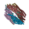
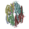
| Entry | Database: PDB / ID: 3zc1 | ||||||
|---|---|---|---|---|---|---|---|
| Title | Crystal structure of AfC3PO | ||||||
 Components Components | AFTRAX | ||||||
 Keywords Keywords |  HYDROLASE / TRANSLIN / TRAX / RNA INTERFERENCE / RNAI / RNA SILENCING / SIRNA / PASSENGER STRAND / RISC HYDROLASE / TRANSLIN / TRAX / RNA INTERFERENCE / RNAI / RNA SILENCING / SIRNA / PASSENGER STRAND / RISC | ||||||
| Function / homology |  Function and homology information Function and homology informationsequence-specific DNA binding / identical protein binding / metal ion bindingSimilarity search - Function | ||||||
| Biological species |   ARCHAEOGLOBUS FULGIDUS (archaea) ARCHAEOGLOBUS FULGIDUS (archaea) | ||||||
| Method | X-RAY DIFFRACTION / SYNCHROTRON / MOLECULAR REPLACEMENT / Resolution: 3.269 Å | ||||||
 Authors Authors | Parizotto, E.A. / Lowe, E.D. / Parker, J.S. | ||||||
 Citation Citation | Journal: Nat.Struct.Mol.Biol. / Year: 2013 Title: Structural Basis for Duplex RNA Recognition and Cleavage by Archaeoglobus Fulgidus C3Po. Authors: Parizotto, E.A. / Lowe, E.D. / Parker, J.S. | ||||||
| History |
|
- Structure visualization
| Structure viewer | Molecule: MolmilJmol/JSmol |
|---|
- Downloads & links
-Download
| PDBx/mmCIF format | 3zc1.cif.gz | 298.6 KB | Display | PDBx/mmCIF format |
|---|---|---|---|---|
| PDB format | pdb3zc1.ent.gz | 241.9 KB | Display | PDB format |
| PDBx/mmJSON format | 3zc1.json.gz | Tree view | PDBx/mmJSON format | |
| Others |  Other downloads Other downloads |
-Validation report
| Arichive directory | https://data.pdbj.org/pub/pdb/validation_reports/zc/3zc1ftp://data.pdbj.org/pub/pdb/validation_reports/zc/3zc1 | HTTPS FTP |
|---|
-Related structure data
| Related structure data | 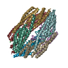 3zc0SC S: Starting model for refinement C: citing same article ( |
|---|---|
| Similar structure data |

-Links
 PDBj
PDBj
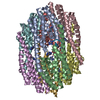
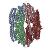
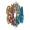
- Assembly
| Deposited unit | 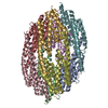
| |||||||||||||||||||||||||||||||||||||||||||||||||||||||||||||||||||||||||||||||
|---|---|---|---|---|---|---|---|---|---|---|---|---|---|---|---|---|---|---|---|---|---|---|---|---|---|---|---|---|---|---|---|---|---|---|---|---|---|---|---|---|---|---|---|---|---|---|---|---|---|---|---|---|---|---|---|---|---|---|---|---|---|---|---|---|---|---|---|---|---|---|---|---|---|---|---|---|---|---|---|---|
| 1 |
| |||||||||||||||||||||||||||||||||||||||||||||||||||||||||||||||||||||||||||||||
| Unit cell |
| |||||||||||||||||||||||||||||||||||||||||||||||||||||||||||||||||||||||||||||||
| Noncrystallographic symmetry (NCS) | NCS domain:
NCS domain segments:
NCS ensembles :
NCS oper:
|
-Components
| #1: Protein | Mass: 22843.406 Da / Num. of mol.: 8 Source method: isolated from a genetically manipulated source Source: (gene. exp.) ARCHAEOGLOBUS FULGIDUS (archaea) / Strain: VC-16Description: DSMZ, GERMAN COLLECTION OF MICROORGANISMS AND CELL CULTURES Plasmid: PTWO-E / Production host:  ESCHERICHIA COLI (E. coli) / Strain (production host): BL21(DE3) / References: UniProt: O28024 ESCHERICHIA COLI (E. coli) / Strain (production host): BL21(DE3) / References: UniProt: O28024#2: Chemical | ChemComp-MG /   Mass: 24.305 Da / Num. of mol.: 5 / Source method: obtained synthetically / Formula: Mg Mass: 24.305 Da / Num. of mol.: 5 / Source method: obtained synthetically / Formula: Mg#3: Water | ChemComp-HOH / | Water Mass: 18.015 Da / Num. of mol.: 8 / Source method: isolated from a natural source / Formula: H2O Mass: 18.015 Da / Num. of mol.: 8 / Source method: isolated from a natural source / Formula: H2OSequence details | CRYSTALLIS | |
|---|
-Experimental details
-Experiment
| Experiment | Method: X-RAY DIFFRACTION / Number of used crystals: 1 |
|---|
- Sample preparation
| Crystal | Density Matthews: 2.6 Å3/Da / Density % sol: 52 % Description: A PRELIMARY MODEL WAS BUILT USING SAD DATA TO 3.41 ANGSTROMS. THIS WAS THEN USED TO SOLVE BY MR A 2.98 ANGSTROM DATASET OF A DIFFERENT CRYSTAL FORM, WHOSE REFINED MODEL WAS THEN USED TO ...Description: A PRELIMARY MODEL WAS BUILT USING SAD DATA TO 3.41 ANGSTROMS. THIS WAS THEN USED TO SOLVE BY MR A 2.98 ANGSTROM DATASET OF A DIFFERENT CRYSTAL FORM, WHOSE REFINED MODEL WAS THEN USED TO SOLVE BY MR THE NATIVE DATASET. |
|---|---|
| Crystal grow | pH: 5.5 Details: 9.5 - 10 % PEG 3K, 100 MM KCL, 200 MM MGCL2, 40 MM SODIUM CACODYLATE PH 5.5, 5 MM DTT. |
-Data collection
| Diffraction | Mean temperature: 100 K |
|---|---|
| Diffraction source | Source: SYNCHROTRON / Site: Diamond  / Beamline: I04 / Wavelength: 0.9763 / Beamline: I04 / Wavelength: 0.9763 |
| Detector | Type: ADSC CCD / Detector: CCD / Date: Oct 1, 2010 / Details: COMPOUND REFRACTIVE LENSES |
| Radiation | Monochromator: DOUBLE CRYSTAL / Protocol: SINGLE WAVELENGTH / Monochromatic (M) / Laue (L): M / Scattering type: x-ray |
| Radiation wavelength | Wavelength: 0.9763 Å / Relative weight: 1 |
| Reflection | Resolution: 3.27→70.75 Å / Num. obs: 29895 / % possible obs: 99.9 % / Redundancy: 9.2 % / Biso Wilson estimate: 106.14 Å2 / Rmerge(I) obs: 0.08 / Net I/σ(I): 17.5 |
| Reflection shell | Resolution: 3.27→3.35 Å / Redundancy: 9.5 % / Rmerge(I) obs: 0.75 / Mean I/σ(I) obs: 2.8 / % possible all: 100 |
- Processing
| Software |
| ||||||||||||||||||||||||||||||||||||||||||||||||||||||||||||||||||||||||||||||||||||||||||||||||||||||||||||||||||||||||||||||||||||||||||||||||||||||||||||||||||||||||||||||||||||||||||||||||||||
|---|---|---|---|---|---|---|---|---|---|---|---|---|---|---|---|---|---|---|---|---|---|---|---|---|---|---|---|---|---|---|---|---|---|---|---|---|---|---|---|---|---|---|---|---|---|---|---|---|---|---|---|---|---|---|---|---|---|---|---|---|---|---|---|---|---|---|---|---|---|---|---|---|---|---|---|---|---|---|---|---|---|---|---|---|---|---|---|---|---|---|---|---|---|---|---|---|---|---|---|---|---|---|---|---|---|---|---|---|---|---|---|---|---|---|---|---|---|---|---|---|---|---|---|---|---|---|---|---|---|---|---|---|---|---|---|---|---|---|---|---|---|---|---|---|---|---|---|---|---|---|---|---|---|---|---|---|---|---|---|---|---|---|---|---|---|---|---|---|---|---|---|---|---|---|---|---|---|---|---|---|---|---|---|---|---|---|---|---|---|---|---|---|---|---|---|---|---|
| Refinement | Method to determine structure: MOLECULAR REPLACEMENT Starting model: PDB ENTRY 3ZC0 Resolution: 3.269→53.56 Å / SU ML: 1.18 / σ(F): 1.35 / Phase error: 31.98 / Stereochemistry target values: ML Details: USED PHENIX.REFINE VERSION 1.7.2. PROTEIN CHAINS COMPLETE EXCEPT 110 OUT OF 1568 SIDE CHAINS MODELLED AS ALANINE STUBS AND 6 TO 9 RESIDUES AT EACH C TERMINUS, MISSING DUE TO INSUFFICIENT ...Details: USED PHENIX.REFINE VERSION 1.7.2. PROTEIN CHAINS COMPLETE EXCEPT 110 OUT OF 1568 SIDE CHAINS MODELLED AS ALANINE STUBS AND 6 TO 9 RESIDUES AT EACH C TERMINUS, MISSING DUE TO INSUFFICIENT ELECTRON DENSITY. MAGNESIUM COORDINATION INTERACTIONS WERE BUILT USING PHENIX.METAL_ COORDINATION.
| ||||||||||||||||||||||||||||||||||||||||||||||||||||||||||||||||||||||||||||||||||||||||||||||||||||||||||||||||||||||||||||||||||||||||||||||||||||||||||||||||||||||||||||||||||||||||||||||||||||
| Solvent computation | Shrinkage radii: 1.11 Å / VDW probe radii: 1.3 Å / Solvent model: FLAT BULK SOLVENT MODEL / Bsol: 76.212 Å2 / ksol: 0.288 e/Å3 | ||||||||||||||||||||||||||||||||||||||||||||||||||||||||||||||||||||||||||||||||||||||||||||||||||||||||||||||||||||||||||||||||||||||||||||||||||||||||||||||||||||||||||||||||||||||||||||||||||||
| Displacement parameters | Biso mean: 103.54 Å2
| ||||||||||||||||||||||||||||||||||||||||||||||||||||||||||||||||||||||||||||||||||||||||||||||||||||||||||||||||||||||||||||||||||||||||||||||||||||||||||||||||||||||||||||||||||||||||||||||||||||
| Refinement step | Cycle: LAST / Resolution: 3.269→53.56 Å
| ||||||||||||||||||||||||||||||||||||||||||||||||||||||||||||||||||||||||||||||||||||||||||||||||||||||||||||||||||||||||||||||||||||||||||||||||||||||||||||||||||||||||||||||||||||||||||||||||||||
| Refine LS restraints |
| ||||||||||||||||||||||||||||||||||||||||||||||||||||||||||||||||||||||||||||||||||||||||||||||||||||||||||||||||||||||||||||||||||||||||||||||||||||||||||||||||||||||||||||||||||||||||||||||||||||
| Refine LS restraints NCS |
| ||||||||||||||||||||||||||||||||||||||||||||||||||||||||||||||||||||||||||||||||||||||||||||||||||||||||||||||||||||||||||||||||||||||||||||||||||||||||||||||||||||||||||||||||||||||||||||||||||||
| LS refinement shell |
|
