 Movie
Movie Controller

[English] 日本語
 Yorodumi
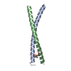
- PDB-3wuv: Structure basis of inactivating cell abscission with chimera peptide 2 -
+ Open data

- Basic information
| Entry | Database: PDB / ID: 3wuv | ||||||
|---|---|---|---|---|---|---|---|
| Title | Structure basis of inactivating cell abscission with chimera peptide 2 | ||||||
 Components Components |
| ||||||
 Keywords Keywords |  CELL CYCLE / Coiled-coil CELL CYCLE / Coiled-coil | ||||||
| Function / homology |  Function and homology information Function and homology informationproteinase activated receptor binding / actomyosin contractile ring assembly / ubiquitin-independent protein catabolic process via the multivesicular body sorting pathway / regulation of extracellular exosome assembly / viral budding / extracellular exosome biogenesis / maintenance of epithelial cell apical/basal polarity / positive regulation of extracellular exosome assembly / regulation of membrane permeability / regulation of centrosome duplication ...proteinase activated receptor binding / actomyosin contractile ring assembly / ubiquitin-independent protein catabolic process via the multivesicular body sorting pathway / regulation of extracellular exosome assembly / viral budding / extracellular exosome biogenesis / maintenance of epithelial cell apical/basal polarity / positive regulation of extracellular exosome assembly / regulation of membrane permeability / regulation of centrosome duplication / cranial skeletal system development / midbody abscission / multivesicular body sorting pathway / bicellular tight junction assembly / actomyosin / positive regulation of exosomal secretion / multivesicular body assembly / Flemming body / RIPK1-mediated regulated necrosis / intercellular bridge / regulation of phosphatidylinositol 3-kinase/protein kinase B signal transduction / viral budding via host ESCRT complex / endoplasmic reticulum exit site / cleavage furrow / Uptake and function of anthrax toxins / mitotic cytokinesis / immunological synapse / bicellular tight junction / centriole / macroautophagy / establishment of protein localization / Budding and maturation of HIV virion / protein homooligomerization / Regulation of necroptotic cell death / calcium-dependent protein binding / extracellular vesicle / melanosome / protein transport / midbody / endosome / focal adhesion / centrosome / apoptotic process / protein homodimerization activity / extracellular exosome / membrane / identical protein binding / cytosol / cytoplasmSimilarity search - Function | ||||||
| Biological species |  Homo sapiens (human) Homo sapiens (human) | ||||||
| Method | X-RAY DIFFRACTION / SYNCHROTRON / MOLECULAR REPLACEMENT / Resolution: 2.79 Å | ||||||
 Authors Authors | Kim, H.J. / Matsuura, A. / Lee, H.H. | ||||||
 Citation Citation | Journal: Proc.Natl.Acad.Sci.USA / Year: 2015 Title: Structural and biochemical insights into the role of testis-expressed gene 14 (TEX14) in forming the stable intercellular bridges of germ cells. Authors: Kim, H.J. / Yoon, J. / Matsuura, A. / Na, J.H. / Lee, W.K. / Kim, H. / Choi, J.W. / Park, J.E. / Park, S.J. / Kim, K.T. / Chang, R. / Lee, B.I. / Yu, Y.G. / Shin, Y.K. / Jeong, C. / Rhee, K. / Lee, H.H. | ||||||
| History |
|
- Structure visualization
| Structure viewer | Molecule: MolmilJmol/JSmol |
|---|
- Downloads & links
-Download
| PDBx/mmCIF format | 3wuv.cif.gz | 163.2 KB | Display | PDBx/mmCIF format |
|---|---|---|---|---|
| PDB format | pdb3wuv.ent.gz | 132.3 KB | Display | PDB format |
| PDBx/mmJSON format | 3wuv.json.gz | Tree view | PDBx/mmJSON format | |
| Others |  Other downloads Other downloads |
-Validation report
| Arichive directory | https://data.pdbj.org/pub/pdb/validation_reports/wu/3wuvftp://data.pdbj.org/pub/pdb/validation_reports/wu/3wuv | HTTPS FTP |
|---|
-Related structure data
| Related structure data |  3wutC  3wuuC  3eirS C: citing same article ( S: Starting model for refinement |
|---|---|
| Similar structure data |


-Links
 PDBj
PDBj


- Assembly
| Deposited unit | 
| ||||||||
|---|---|---|---|---|---|---|---|---|---|
| 1 | 
| ||||||||
| 2 | 
| ||||||||
| 3 | 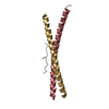
| ||||||||
| 4 | 
| ||||||||
| 5 | 
| ||||||||
| 6 | 
| ||||||||
| Unit cell |
|
-Components
| #1: Protein | Centrosome / Cep55 / Up-regulated in colon cancer 6 Mass: 7243.210 Da / Num. of mol.: 12 / Fragment: UNP residues 160-217 Source method: isolated from a genetically manipulated source Source: (gene. exp.) Homo sapiens (human) / Gene: CEP55 / Plasmid: pGST2 / Production host:  Escherichia coli BL21(DE3) (bacteria) / References: UniProt: Q53EZ4 Escherichia coli BL21(DE3) (bacteria) / References: UniProt: Q53EZ4#2: Protein/peptide | / ALG-2-interacting protein X / ALIXMass: 1543.673 Da / Num. of mol.: 6 / Mutation: P796D/P807I/G808P/Y809P / Source method: obtained synthetically / Details: synthetic peptide / Source: (synth.) Homo sapiens (human) / References: UniProt: Q8WUM4#3: Water | ChemComp-HOH / | Water Mass: 18.015 Da / Num. of mol.: 170 / Source method: isolated from a natural source / Formula: H2O Mass: 18.015 Da / Num. of mol.: 170 / Source method: isolated from a natural source / Formula: H2O |
|---|
-Experimental details
-Experiment
| Experiment | Method: X-RAY DIFFRACTION / Number of used crystals: 1 |
|---|
- Sample preparation
| Crystal | Density Matthews: 7.21 Å3/Da / Density % sol: 82.93 % |
|---|---|
| Crystal grow | Temperature: 295 K / Method: vapor diffusion, hanging drop / pH: 5 Details: 0.8M ammonium sulfate, pH 5.0, VAPOR DIFFUSION, HANGING DROP, temperature 295K |
-Data collection
| Diffraction | Mean temperature: 100 K |
|---|---|
| Diffraction source | Source: SYNCHROTRON / Site: PAL/PLS  / Beamline: 5C (4A) / Wavelength: 1 Å / Beamline: 5C (4A) / Wavelength: 1 Å |
| Detector | Type: ADSC QUANTUM 315r / Detector: CCD / Date: Apr 24, 2014 |
| Radiation | Monochromator: si 111 DCM / Protocol: SINGLE WAVELENGTH / Monochromatic (M) / Laue (L): M / Scattering type: x-ray |
| Radiation wavelength | Wavelength: 1 Å / Relative weight: 1 |
| Reflection | Resolution: 2.79→50 Å / Num. all: 67538 / Num. obs: 67471 / % possible obs: 99.9 % / Observed criterion σ(I): -3 / Biso Wilson estimate: 46.75 Å2 |
| Reflection shell | Highest resolution: 2.8 Å / % possible all: 99.9 |
- Processing
| Software |
| |||||||||||||||||||||||||||||||||||||||||||||||||||||||||||||||||||||||||||||||||||||||||||||||||||||||||
|---|---|---|---|---|---|---|---|---|---|---|---|---|---|---|---|---|---|---|---|---|---|---|---|---|---|---|---|---|---|---|---|---|---|---|---|---|---|---|---|---|---|---|---|---|---|---|---|---|---|---|---|---|---|---|---|---|---|---|---|---|---|---|---|---|---|---|---|---|---|---|---|---|---|---|---|---|---|---|---|---|---|---|---|---|---|---|---|---|---|---|---|---|---|---|---|---|---|---|---|---|---|---|---|---|---|---|
| Refinement | Method to determine structure: MOLECULAR REPLACEMENT Starting model: 3EIR Resolution: 2.79→48.644 Å / FOM work R set: 0.8209 / SU ML: 0.24 / σ(F): 1.34 / Phase error: 24.68 / Stereochemistry target values: ML
| |||||||||||||||||||||||||||||||||||||||||||||||||||||||||||||||||||||||||||||||||||||||||||||||||||||||||
| Solvent computation | Shrinkage radii: 0.9 Å / VDW probe radii: 1.11 Å / Solvent model: FLAT BULK SOLVENT MODEL | |||||||||||||||||||||||||||||||||||||||||||||||||||||||||||||||||||||||||||||||||||||||||||||||||||||||||
| Displacement parameters | Biso max: 198.22 Å2 / Biso mean: 62.97 Å2 / Biso min: 14.16 Å2 | |||||||||||||||||||||||||||||||||||||||||||||||||||||||||||||||||||||||||||||||||||||||||||||||||||||||||
| Refinement step | Cycle: LAST / Resolution: 2.79→48.644 Å
| |||||||||||||||||||||||||||||||||||||||||||||||||||||||||||||||||||||||||||||||||||||||||||||||||||||||||
| Refine LS restraints |
| |||||||||||||||||||||||||||||||||||||||||||||||||||||||||||||||||||||||||||||||||||||||||||||||||||||||||
| LS refinement shell | Refine-ID: X-RAY DIFFRACTION / Total num. of bins used: 14
|
