[English] 日本語
Yorodumi
- PDB-3wus: Crystal Structure of the Vif-Binding Domain of Human APOBEC3F -

+
Open data


ID or keywords:

Loading...

-
Basic information

Entry
Database: PDB / ID: 3wus
TitleCrystal Structure of the Vif-Binding Domain of Human APOBEC3F
ComponentsDNA dC->dU-editing enzyme APOBEC-3F
KeywordsHYDROLASE / APOBEC3F / A3F / Zinc binding / antiviral enzyme / CYTIDINE DEAMINASE / DNA binding / HIV-1 VIF / single-stranded polynucleotide
Function / homology
Function and homology information


apolipoprotein B mRNA editing enzyme complex / base conversion or substitution editing / single-stranded DNA cytosine deaminase / DNA cytosine deamination / cytidine to uridine editing / : / cytidine deaminase activity / clearance of foreign intracellular DNA / negative regulation of single stranded viral RNA replication via double stranded DNA intermediate / negative regulation of viral process ...apolipoprotein B mRNA editing enzyme complex / base conversion or substitution editing / single-stranded DNA cytosine deaminase / DNA cytosine deamination / cytidine to uridine editing / : / cytidine deaminase activity / clearance of foreign intracellular DNA / negative regulation of single stranded viral RNA replication via double stranded DNA intermediate / negative regulation of viral process / retrotransposon silencing / : / negative regulation of viral genome replication / positive regulation of defense response to virus by host / P-body / defense response to virus / ribonucleoprotein complex / innate immune response / RNA binding / zinc ion binding / identical protein binding / nucleus / cytoplasm
Similarity search - Function
APOBEC-like N-terminal domain / Novel AID APOBEC clade 2 / APOBEC/CMP deaminase, zinc-binding / Cytidine and deoxycytidylate deaminases zinc-binding region signature. / Cytidine and deoxycytidylate deaminase domain / Cytidine and deoxycytidylate deaminases domain profile. / Cytidine deaminase-like
Similarity search - Domain/homology
DNA dC->dU-editing enzyme APOBEC-3F
Similarity search - Component
Biological speciesHomo sapiens (human)
MethodX-RAY DIFFRACTION / SYNCHROTRON / MOLECULAR REPLACEMENT / Resolution: 2.54 Å
AuthorsNakashima, M. / Kawamura, T. / Ode, H. / Watanabe, N. / Iwatani, Y.
CitationJournal: J.Virol. / Year: 2015
Title: Structural Insights into HIV-1 Vif-APOBEC3F Interaction.
Authors: Nakashima, M. / Ode, H. / Kawamura, T. / Kitamura, S. / Naganawa, Y. / Awazu, H. / Tsuzuki, S. / Matsuoka, K. / Nemoto, M. / Hachiya, A. / Sugiura, W. / Yokomaku, Y. / Watanabe, N. / Iwatani, Y.
History
DepositionMay 2, 2014Deposition site: PDBJ / Processing site: PDBJ
Revision 1.0Jun 24, 2015Provider: repository / Type: Initial release
Revision 1.1Jan 27, 2016Group: Database references
Revision 1.2Nov 22, 2017Group: Refinement description / Category: software / Item: _software.name
Revision 1.3Nov 8, 2023Group: Data collection / Database references ...Data collection / Database references / Derived calculations / Refinement description
Category: chem_comp_atom / chem_comp_bond ...chem_comp_atom / chem_comp_bond / database_2 / pdbx_initial_refinement_model / pdbx_struct_conn_angle / struct_conn / struct_ref_seq_dif / struct_site
Item: _database_2.pdbx_DOI / _database_2.pdbx_database_accession ..._database_2.pdbx_DOI / _database_2.pdbx_database_accession / _pdbx_struct_conn_angle.ptnr1_auth_comp_id / _pdbx_struct_conn_angle.ptnr1_auth_seq_id / _pdbx_struct_conn_angle.ptnr1_label_atom_id / _pdbx_struct_conn_angle.ptnr1_label_comp_id / _pdbx_struct_conn_angle.ptnr1_label_seq_id / _pdbx_struct_conn_angle.ptnr3_auth_comp_id / _pdbx_struct_conn_angle.ptnr3_auth_seq_id / _pdbx_struct_conn_angle.ptnr3_label_atom_id / _pdbx_struct_conn_angle.ptnr3_label_comp_id / _pdbx_struct_conn_angle.ptnr3_label_seq_id / _pdbx_struct_conn_angle.value / _struct_conn.pdbx_dist_value / _struct_conn.ptnr1_auth_asym_id / _struct_conn.ptnr1_auth_comp_id / _struct_conn.ptnr1_auth_seq_id / _struct_conn.ptnr1_label_asym_id / _struct_conn.ptnr1_label_atom_id / _struct_conn.ptnr1_label_comp_id / _struct_conn.ptnr1_label_seq_id / _struct_conn.ptnr2_auth_asym_id / _struct_conn.ptnr2_auth_comp_id / _struct_conn.ptnr2_auth_seq_id / _struct_conn.ptnr2_label_asym_id / _struct_conn.ptnr2_label_atom_id / _struct_conn.ptnr2_label_comp_id / _struct_ref_seq_dif.details / _struct_site.pdbx_auth_asym_id / _struct_site.pdbx_auth_comp_id / _struct_site.pdbx_auth_seq_id

-
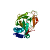
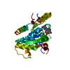
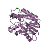
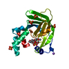
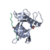
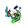
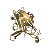
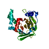
Structure visualization

Structure viewerMolecule:
MolmilJmol/JSmol

Downloads & links

-
Assembly

Deposited unit
A: DNA dC->dU-editing enzyme APOBEC-3F
B: DNA dC->dU-editing enzyme APOBEC-3F
hetero molecules


Theoretical massNumber of molelcules
Total (without water)45,8684
Polymers45,7382
Non-polymers1312
Water1267
1
A: DNA dC->dU-editing enzyme APOBEC-3F
hetero molecules


Theoretical massNumber of molelcules
Total (without water)22,9342
Polymers22,8691
Non-polymers651
Water181
TypeNameSymmetry operationNumber
identity operation1_555x,y,z1
MethodPISA
2
B: DNA dC->dU-editing enzyme APOBEC-3F
hetero molecules


Theoretical massNumber of molelcules
Total (without water)22,9342
Polymers22,8691
Non-polymers651
Water181
TypeNameSymmetry operationNumber
identity operation1_555x,y,z1
MethodPISA
Unit cell
Length a, b, c (Å)117.294, 117.294, 78.960
Angle α, β, γ (deg.)90.00, 90.00, 120.00
Int Tables number152
Space group name H-MP3121

-
Components

#1: Protein DNA dC->dU-editing enzyme APOBEC-3F / Apolipoprotein B mRNA-editing enzyme catalytic polypeptide-like 3F / A3F


Mass: 22868.834 Da / Num. of mol.: 2 / Fragment: C-TERMINAL DOMAIN, UNP RESIDUES 187-373
Source method: isolated from a genetically manipulated source
Source: (gene. exp.) Homo sapiens (human) / Gene: APOBEC3F / Plasmid: PET41A(+) / Production host: Escherichia coli (E. coli) / Strain (production host): ROSETTA2(DE3)PLYSS
References: UniProt: Q8IUX4, Hydrolases; Acting on carbon-nitrogen bonds, other than peptide bonds; In cyclic amidines
#2: Chemical ChemComp-ZN / ZINC ION


Mass: 65.409 Da / Num. of mol.: 2 / Source method: obtained synthetically / Formula: Zn
#3: Water ChemComp-HOH / water / Water


Mass: 18.015 Da / Num. of mol.: 7 / Source method: isolated from a natural source / Formula: H2O

-
Experimental details

-
Experiment

ExperimentMethod: X-RAY DIFFRACTION / Number of used crystals: 1

-
Sample preparation

CrystalDensity Matthews: 3.43 Å3/Da / Density % sol: 64.12 %
Crystal growTemperature: 293 K / Method: vapor diffusion, hanging drop / pH: 9
Details: 8.5% PEG 20000, 2% Dioxane, 300mM L-Arginine HCl, 85mM Na BICINE, pH 9, VAPOR DIFFUSION, HANGING DROP, temperature 293K

-
Data collection

DiffractionMean temperature: 95 K
Diffraction sourceSource: SYNCHROTRON / Site: Photon Factory / Beamline: BL-5A / Wavelength: 1 Å
DetectorType: ADSC QUANTUM 315r / Detector: CCD / Date: Dec 16, 2012
RadiationMonochromator: Si(111) double crystal monochromator / Protocol: SINGLE WAVELENGTH / Monochromatic (M) / Laue (L): M / Scattering type: x-ray
Radiation wavelengthWavelength: 1 Å / Relative weight: 1
ReflectionResolution: 2.54→101.58 Å / Num. obs: 20935 / % possible obs: 99.9 % / Redundancy: 10.9 % / Rsym value: 0.107 / Net I/σ(I): 36.1
Reflection shellResolution: 2.54→2.58 Å / % possible all: 98.7

-
Processing

Software
NameVersionClassification
SERGUIdata collection
PHASERphasing
REFMAC5.8.0069refinement
HKL-2000data reduction
HKL-2000data scaling
RefinementMethod to determine structure: MOLECULAR REPLACEMENT
Starting model: PDB ENTRY 3VOW
Resolution: 2.54→50 Å / Cor.coef. Fo:Fc: 0.948 / Cor.coef. Fo:Fc free: 0.912 / SU B: 9.709 / SU ML: 0.205 / Cross valid method: THROUGHOUT / ESU R: 0.34 / ESU R Free: 0.264 / Stereochemistry target values: MAXIMUM LIKELIHOOD / Details: HYDROGENS HAVE BEEN ADDED IN THE RIDING POSITIONS
RfactorNum. reflection% reflectionSelection details
Rfree0.25648 1069 5.1 %RANDOM
Rwork0.19979 ---
obs0.20266 19812 99.78 %-
Solvent computationIon probe radii: 0.8 Å / Shrinkage radii: 0.8 Å / VDW probe radii: 1.2 Å / Solvent model: MASK
Displacement parametersBiso mean: 54.789 Å2
Baniso -1Baniso -2Baniso -3
1-1.39 Å20.69 Å20 Å2
2--1.39 Å2-0 Å2
3----4.5 Å2
Refinement stepCycle: LAST / Resolution: 2.54→50 Å
ProteinNucleic acidLigandSolventTotal
Num. atoms3130 0 2 7 3139
Refine LS restraints
Refine-IDTypeDev idealDev ideal targetNumber
X-RAY DIFFRACTIONr_bond_refined_d0.0160.0193248
X-RAY DIFFRACTIONr_bond_other_d0.0010.022832
X-RAY DIFFRACTIONr_angle_refined_deg1.8631.9114416
X-RAY DIFFRACTIONr_angle_other_deg0.89536522
X-RAY DIFFRACTIONr_dihedral_angle_1_deg8.535366
X-RAY DIFFRACTIONr_dihedral_angle_2_deg34.51523.556180
X-RAY DIFFRACTIONr_dihedral_angle_3_deg19.56715506
X-RAY DIFFRACTIONr_dihedral_angle_4_deg17.9651516
X-RAY DIFFRACTIONr_chiral_restr0.1070.2428
X-RAY DIFFRACTIONr_gen_planes_refined0.0080.0213694
X-RAY DIFFRACTIONr_gen_planes_other0.0010.02870
X-RAY DIFFRACTIONr_nbd_refined
X-RAY DIFFRACTIONr_nbd_other
X-RAY DIFFRACTIONr_nbtor_refined
X-RAY DIFFRACTIONr_nbtor_other
X-RAY DIFFRACTIONr_xyhbond_nbd_refined
X-RAY DIFFRACTIONr_xyhbond_nbd_other
X-RAY DIFFRACTIONr_metal_ion_refined
X-RAY DIFFRACTIONr_metal_ion_other
X-RAY DIFFRACTIONr_symmetry_vdw_refined
X-RAY DIFFRACTIONr_symmetry_vdw_other
X-RAY DIFFRACTIONr_symmetry_hbond_refined
X-RAY DIFFRACTIONr_symmetry_hbond_other
X-RAY DIFFRACTIONr_symmetry_metal_ion_refined
X-RAY DIFFRACTIONr_symmetry_metal_ion_other
X-RAY DIFFRACTIONr_mcbond_it4.5655.1751470
X-RAY DIFFRACTIONr_mcbond_other4.5625.1721469
X-RAY DIFFRACTIONr_mcangle_it7.0227.7521834
X-RAY DIFFRACTIONr_mcangle_other7.0237.7561835
X-RAY DIFFRACTIONr_scbond_it5.3015.7141778
X-RAY DIFFRACTIONr_scbond_other5.2935.7071775
X-RAY DIFFRACTIONr_scangle_it
X-RAY DIFFRACTIONr_scangle_other8.2118.372581
X-RAY DIFFRACTIONr_long_range_B_refined11.13542.5043793
X-RAY DIFFRACTIONr_long_range_B_other11.13442.5143793
X-RAY DIFFRACTIONr_rigid_bond_restr
X-RAY DIFFRACTIONr_sphericity_free
X-RAY DIFFRACTIONr_sphericity_bonded
LS refinement shellResolution: 2.544→2.61 Å / Total num. of bins used: 20
RfactorNum. reflection% reflection
Rfree0.359 57 -
Rwork0.315 1454 -
obs--98.37 %

+
About Yorodumi

-
News

-
Feb 9, 2022. New format data for meta-information of EMDB entries

New format data for meta-information of EMDB entries

  • Version 3 of the EMDB header file is now the official format.
  • The previous official version 1.9 will be removed from the archive.

Related info.:EMDB header

External links:wwPDB to switch to version 3 of the EMDB data model

-
Aug 12, 2020. Covid-19 info

Covid-19 info

URL: https://pdbj.org/emnavi/covid19.php

New page: Covid-19 featured information page in EM Navigator.

Related info.:Covid-19 info / Mar 5, 2020. Novel coronavirus structure data

+
Mar 5, 2020. Novel coronavirus structure data

Novel coronavirus structure data

Related info.:Yorodumi Speices / Aug 12, 2020. Covid-19 info

External links:COVID-19 featured content - PDBj / Molecule of the Month (242):Coronavirus Proteases

+
Jan 31, 2019. EMDB accession codes are about to change! (news from PDBe EMDB page)

EMDB accession codes are about to change! (news from PDBe EMDB page)

  • The allocation of 4 digits for EMDB accession codes will soon come to an end. Whilst these codes will remain in use, new EMDB accession codes will include an additional digit and will expand incrementally as the available range of codes is exhausted. The current 4-digit format prefixed with “EMD-” (i.e. EMD-XXXX) will advance to a 5-digit format (i.e. EMD-XXXXX), and so on. It is currently estimated that the 4-digit codes will be depleted around Spring 2019, at which point the 5-digit format will come into force.
  • The EM Navigator/Yorodumi systems omit the EMD- prefix.

Related info.:Q: What is EMD? / ID/Accession-code notation in Yorodumi/EM Navigator

External links:EMDB Accession Codes are Changing Soon! / Contact to PDBj

+
Jul 12, 2017. Major update of PDB

Major update of PDB

  • wwPDB released updated PDB data conforming to the new PDBx/mmCIF dictionary.
  • This is a major update changing the version number from 4 to 5, and with Remediation, in which all the entries are updated.
  • In this update, many items about electron microscopy experimental information are reorganized (e.g. em_software).
  • Now, EM Navigator and Yorodumi are based on the updated data.

External links:wwPDB Remediation / Enriched Model Files Conforming to OneDep Data Standards Now Available in the PDB FTP Archive

-
Yorodumi

Thousand views of thousand structures

  • Yorodumi is a browser for structure data from EMDB, PDB, SASBDB, etc.
  • This page is also the successor to EM Navigator detail page, and also detail information page/front-end page for Omokage search.
  • The word "yorodu" (or yorozu) is an old Japanese word meaning "ten thousand". "mi" (miru) is to see.

Related info.:EMDB / PDB / SASBDB / Comparison of 3 databanks / Yorodumi Search / Aug 31, 2016. New EM Navigator & Yorodumi / Yorodumi Papers / Jmol/JSmol / Function and homology information / Changes in new EM Navigator and Yorodumi

Read more