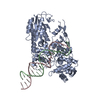
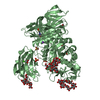
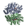
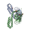
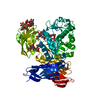
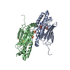
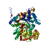
[English] 日本語
Yorodumi
- PDB-3wnk: Crystal Structure of Bacillus circulans T-3040 cycloisomaltooligo... -

+
Open data


ID or keywords:

Loading...

-
Basic information

Entry
Database: PDB / ID: 3wnk
TitleCrystal Structure of Bacillus circulans T-3040 cycloisomaltooligosaccharide glucanotransferase
ComponentsCycloisomaltooligosaccharide glucanotransferase
KeywordsTRANSFERASE / C2 type immunoglobulin fold / (beta/alpha)8-barrel / beta-jelly roll / Greek key / Glycoside Hydrolase / alpha-1 / 6-glucan
Function / homology
Function and homology information


cycloisomaltooligosaccharide glucanotransferase / glycosyltransferase activity / carbohydrate binding
Similarity search - Function
Glycosyl hydrolase family 66 / Glycosyl hydrolase family 66 / Carbohydrate binding module (family 35) / Carbohydrate binding module (family 6) / CBM6 (carbohydrate binding type-6) domain profile. / Carbohydrate binding module family 6 / Galactose-binding domain-like / Golgi alpha-mannosidase II / Glycosyl hydrolase, all-beta / Galactose-binding-like domain superfamily ...Glycosyl hydrolase family 66 / Glycosyl hydrolase family 66 / Carbohydrate binding module (family 35) / Carbohydrate binding module (family 6) / CBM6 (carbohydrate binding type-6) domain profile. / Carbohydrate binding module family 6 / Galactose-binding domain-like / Golgi alpha-mannosidase II / Glycosyl hydrolase, all-beta / Galactose-binding-like domain superfamily / Glycosidases / Jelly Rolls / Immunoglobulins / TIM Barrel / Alpha-Beta Barrel / Immunoglobulin-like fold / Immunoglobulin-like / Sandwich / Mainly Beta / Alpha Beta
Similarity search - Domain/homology
ACETATE ION / : / Cycloisomaltooligosaccharide glucanotransferase
Similarity search - Component
Biological speciesBacillus circulans (bacteria)
MethodX-RAY DIFFRACTION / SYNCHROTRON / MAD / Resolution: 2.3 Å
AuthorsSuzuki, N. / Fujimoto, Z. / Kim, Y.M. / Momma, M. / Kishine, N. / Suzuki, R. / Kobayashi, M. / Kimura, A. / Funane, K.
Citation
Journal: J.Biol.Chem. / Year: 2014
Title: Structural elucidation of the cyclization mechanism of alpha-1,6-glucan by Bacillus circulans T-3040 cycloisomaltooligosaccharide glucanotransferase.
Authors: Suzuki, N. / Fujimoto, Z. / Kim, Y.M. / Momma, M. / Kishine, N. / Suzuki, R. / Suzuki, S. / Kitamura, S. / Kobayashi, M. / Kimura, A. / Funane, K.
#1: Journal: Acta Crystallogr.,Sect.F / Year: 2013
Title: Crystallization and preliminary X-ray crystallographic analysis of cycloisomaltooligosaccharide glucanotransferase from Bacillus circulans T-3040.
Authors: Suzuki, N. / Kim, Y.M. / Momma, M. / Fujimoto, Z. / Kobayashi, M. / Kimura, A. / Funane, K.
#2: Journal: Biochim.Biophys.Acta / Year: 2011
Title: Deletion analysis of regions at the C-terminal part of cycloisomaltooligosaccharide glucanotransferase from Bacillus circulans T-3040.
Authors: Funane, K. / Kawabata, Y. / Suzuki, R. / Kim, Y.M. / Kang, H.K. / Suzuki, N. / Fujimoto, Z. / Kimura, A. / Kobayashi, M.
History
DepositionDec 10, 2013Deposition site: PDBJ / Processing site: PDBJ
Revision 1.0Feb 5, 2014Provider: repository / Type: Initial release
Revision 1.1Mar 4, 2015Group: Database references
Revision 1.2Mar 20, 2024Group: Data collection / Database references / Derived calculations
Category: chem_comp_atom / chem_comp_bond ...chem_comp_atom / chem_comp_bond / database_2 / pdbx_struct_conn_angle / struct_conn / struct_ref_seq_dif / struct_site
Item: _database_2.pdbx_DOI / _database_2.pdbx_database_accession ..._database_2.pdbx_DOI / _database_2.pdbx_database_accession / _pdbx_struct_conn_angle.ptnr1_auth_comp_id / _pdbx_struct_conn_angle.ptnr1_auth_seq_id / _pdbx_struct_conn_angle.ptnr1_label_alt_id / _pdbx_struct_conn_angle.ptnr1_label_asym_id / _pdbx_struct_conn_angle.ptnr1_label_atom_id / _pdbx_struct_conn_angle.ptnr1_label_comp_id / _pdbx_struct_conn_angle.ptnr1_label_seq_id / _pdbx_struct_conn_angle.ptnr2_auth_comp_id / _pdbx_struct_conn_angle.ptnr2_auth_seq_id / _pdbx_struct_conn_angle.ptnr2_label_asym_id / _pdbx_struct_conn_angle.ptnr2_label_atom_id / _pdbx_struct_conn_angle.ptnr2_label_comp_id / _pdbx_struct_conn_angle.ptnr3_auth_comp_id / _pdbx_struct_conn_angle.ptnr3_auth_seq_id / _pdbx_struct_conn_angle.ptnr3_label_asym_id / _pdbx_struct_conn_angle.ptnr3_label_atom_id / _pdbx_struct_conn_angle.ptnr3_label_comp_id / _pdbx_struct_conn_angle.ptnr3_label_seq_id / _pdbx_struct_conn_angle.value / _struct_conn.pdbx_dist_value / _struct_conn.pdbx_ptnr1_label_alt_id / _struct_conn.ptnr1_auth_comp_id / _struct_conn.ptnr1_auth_seq_id / _struct_conn.ptnr1_label_asym_id / _struct_conn.ptnr1_label_atom_id / _struct_conn.ptnr1_label_comp_id / _struct_conn.ptnr1_label_seq_id / _struct_conn.ptnr2_auth_comp_id / _struct_conn.ptnr2_auth_seq_id / _struct_conn.ptnr2_label_asym_id / _struct_conn.ptnr2_label_atom_id / _struct_conn.ptnr2_label_comp_id / _struct_ref_seq_dif.details / _struct_site.pdbx_auth_asym_id / _struct_site.pdbx_auth_comp_id / _struct_site.pdbx_auth_seq_id

-
Structure visualization

Structure viewerMolecule:
MolmilJmol/JSmol

Downloads & links

-
Assembly

Deposited unit
A: Cycloisomaltooligosaccharide glucanotransferase
hetero molecules


Theoretical massNumber of molelcules
Total (without water)81,77419
Polymers80,2211
Non-polymers1,55318
Water9,890549
1


  • Idetical with deposited unit
  • defined by author&software
TypeNameSymmetry operationNumber
identity operation1_555x,y,z1
MethodPISA
Unit cell
Length a, b, c (Å)106.437, 106.437, 160.625
Angle α, β, γ (deg.)90.00, 90.00, 120.00
Int Tables number152
Space group name H-MP3121
Components on special symmetry positions
IDModelComponents
11A-1179-

HOH

21A-1314-

HOH

-
Components

-
Protein , 1 types, 1 molecules A

#1: Protein Cycloisomaltooligosaccharide glucanotransferase / / CITase


Mass: 80221.305 Da / Num. of mol.: 1 / Fragment: UNP RESIDUES 39-738
Source method: isolated from a genetically manipulated source
Source: (gene. exp.) Bacillus circulans (bacteria) / Strain: T-3040 / Gene: cit / Plasmid: pET-28a / Production host: Escherichia coli (E. coli) / Strain (production host): BL21(DE3)
References: UniProt: P94286, cycloisomaltooligosaccharide glucanotransferase

-
Non-polymers , 6 types, 567 molecules

#2: Chemical
ChemComp-CD / CADMIUM ION


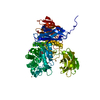
Mass: 112.411 Da / Num. of mol.: 10 / Source method: obtained synthetically / Formula: Cd
#3: Chemical ChemComp-CA / CALCIUM ION


Mass: 40.078 Da / Num. of mol.: 2 / Source method: obtained synthetically / Formula: Ca
#4: Chemical ChemComp-NA / SODIUM ION


Mass: 22.990 Da / Num. of mol.: 2 / Source method: obtained synthetically / Formula: Na
#5: Chemical ChemComp-ACT / ACETATE ION / Acetate


Mass: 59.044 Da / Num. of mol.: 2 / Source method: obtained synthetically / Formula: C2H3O2
#6: Chemical ChemComp-GOL / GLYCEROL / GLYCERIN / PROPANE-1,2,3-TRIOL / Glycerol


Mass: 92.094 Da / Num. of mol.: 2 / Source method: obtained synthetically / Formula: C3H8O3
#7: Water ChemComp-HOH / water / Water


Mass: 18.015 Da / Num. of mol.: 549 / Source method: isolated from a natural source / Formula: H2O

-
Details

Sequence detailsTHE DEPOSITORS CONFIRM THAT RESIDUE 278 IS PHE AND THAT SWISSPROT IS INCORRECT AT THIS POSITION.

-
Experimental details

-
Experiment

ExperimentMethod: X-RAY DIFFRACTION / Number of used crystals: 1

-
Sample preparation

CrystalDensity Matthews: 3.27 Å3/Da / Density % sol: 62.43 %
Crystal growTemperature: 293 K / Method: vapor diffusion, sitting drop / pH: 7.5
Details: 1.0M sodium acetate trihydrate, 0.1M HEPES, 0.05M cadmium sulfate hydrate, 12%(v/v) glycerol, pH 7.5, VAPOR DIFFUSION, SITTING DROP, temperature 293K

-
Data collection

DiffractionMean temperature: 95 K
Diffraction sourceSource: SYNCHROTRON / Site: Photon Factory / Beamline: BL-6A / Wavelength: 0.97800, 0.97885, 0.97931, 0.98319, 0.96408
DetectorType: ADSC QUANTUM 4r / Detector: CCD / Date: May 23, 2007 / Details: 1m long bent plane mirror of fused quartz
RadiationMonochromator: Triangular Si(111) / Protocol: MAD / Monochromatic (M) / Laue (L): M / Scattering type: x-ray
Radiation wavelength
IDWavelength (Å)Relative weight
10.9781
20.978851
30.979311
40.983191
50.964081
ReflectionResolution: 2.3→44.34 Å / Num. all: 47422 / Num. obs: 47410 / % possible obs: 100 % / Observed criterion σ(F): 0 / Observed criterion σ(I): -3 / Redundancy: 16.6 % / Biso Wilson estimate: 29.9 Å2 / Rsym value: 0.079 / Net I/σ(I): 47.1
Reflection shellResolution: 2.3→2.38 Å / Redundancy: 16.7 % / Mean I/σ(I) obs: 10.6 / Num. unique all: 4667 / Rsym value: 0.423 / % possible all: 100

-
Processing

Software
NameVersionClassification
ADSCQuantumdata collection
SOLVEphasing
REFMAC5.7.0029refinement
HKL-2000data reduction
SCALEPACKdata scaling
RefinementMethod to determine structure: MAD / Resolution: 2.3→44.34 Å / Cor.coef. Fo:Fc: 0.966 / Cor.coef. Fo:Fc free: 0.954 / SU B: 4.473 / SU ML: 0.109 / Cross valid method: THROUGHOUT / ESU R: 0.204 / ESU R Free: 0.167 / Stereochemistry target values: MAXIMUM LIKELIHOOD
RfactorNum. reflection% reflectionSelection details
Rfree0.19203 2396 5.1 %RANDOM
Rwork0.15764 ---
obs0.15938 44946 99.91 %-
Solvent computationIon probe radii: 0.8 Å / Shrinkage radii: 0.8 Å / VDW probe radii: 1.2 Å / Solvent model: MASK
Displacement parametersBiso mean: 37.707 Å2
Baniso -1Baniso -2Baniso -3
1-1.04 Å21.04 Å20 Å2
2--1.04 Å20 Å2
3----3.39 Å2
Refine analyzeLuzzati coordinate error obs: 0.109 Å
Refinement stepCycle: LAST / Resolution: 2.3→44.34 Å
ProteinNucleic acidLigandSolventTotal
Num. atoms5591 0 34 549 6174
Refine LS restraints
Refine-IDTypeDev idealDev ideal targetNumber
X-RAY DIFFRACTIONr_bond_refined_d0.0080.0195762
X-RAY DIFFRACTIONr_bond_other_d0.0010.025070
X-RAY DIFFRACTIONr_angle_refined_deg1.2351.9177840
X-RAY DIFFRACTIONr_angle_other_deg0.724311627
X-RAY DIFFRACTIONr_dihedral_angle_1_deg6.5165711
X-RAY DIFFRACTIONr_dihedral_angle_2_deg36.35924.716299
X-RAY DIFFRACTIONr_dihedral_angle_3_deg13.18615864
X-RAY DIFFRACTIONr_dihedral_angle_4_deg16.861525
X-RAY DIFFRACTIONr_chiral_restr0.0760.2829
X-RAY DIFFRACTIONr_gen_planes_refined0.0050.026803
X-RAY DIFFRACTIONr_gen_planes_other0.0010.021460
X-RAY DIFFRACTIONr_nbd_refined
X-RAY DIFFRACTIONr_nbd_other
X-RAY DIFFRACTIONr_nbtor_refined
X-RAY DIFFRACTIONr_nbtor_other
X-RAY DIFFRACTIONr_xyhbond_nbd_refined
X-RAY DIFFRACTIONr_xyhbond_nbd_other
X-RAY DIFFRACTIONr_metal_ion_refined
X-RAY DIFFRACTIONr_metal_ion_other
X-RAY DIFFRACTIONr_symmetry_vdw_refined
X-RAY DIFFRACTIONr_symmetry_vdw_other
X-RAY DIFFRACTIONr_symmetry_hbond_refined
X-RAY DIFFRACTIONr_symmetry_hbond_other
X-RAY DIFFRACTIONr_symmetry_metal_ion_refined
X-RAY DIFFRACTIONr_symmetry_metal_ion_other
X-RAY DIFFRACTIONr_mcbond_it
X-RAY DIFFRACTIONr_mcbond_other
X-RAY DIFFRACTIONr_mcangle_it
X-RAY DIFFRACTIONr_mcangle_other
X-RAY DIFFRACTIONr_scbond_it
X-RAY DIFFRACTIONr_scbond_other
X-RAY DIFFRACTIONr_scangle_it
X-RAY DIFFRACTIONr_scangle_other
X-RAY DIFFRACTIONr_long_range_B_refined
X-RAY DIFFRACTIONr_long_range_B_other
X-RAY DIFFRACTIONr_rigid_bond_restr
X-RAY DIFFRACTIONr_sphericity_free
X-RAY DIFFRACTIONr_sphericity_bonded
LS refinement shellResolution: 2.3→2.36 Å / Total num. of bins used: 20
RfactorNum. reflection% reflection
Rfree0.269 175 -
Rwork0.205 3280 -
obs--99.8 %

+
About Yorodumi

-
News

-
Feb 9, 2022. New format data for meta-information of EMDB entries

New format data for meta-information of EMDB entries

  • Version 3 of the EMDB header file is now the official format.
  • The previous official version 1.9 will be removed from the archive.

Related info.:EMDB header

External links:wwPDB to switch to version 3 of the EMDB data model

-
Aug 12, 2020. Covid-19 info

Covid-19 info

URL: https://pdbj.org/emnavi/covid19.php

New page: Covid-19 featured information page in EM Navigator.

Related info.:Covid-19 info / Mar 5, 2020. Novel coronavirus structure data

+
Mar 5, 2020. Novel coronavirus structure data

Novel coronavirus structure data

Related info.:Yorodumi Speices / Aug 12, 2020. Covid-19 info

External links:COVID-19 featured content - PDBj / Molecule of the Month (242):Coronavirus Proteases

+
Jan 31, 2019. EMDB accession codes are about to change! (news from PDBe EMDB page)

EMDB accession codes are about to change! (news from PDBe EMDB page)

  • The allocation of 4 digits for EMDB accession codes will soon come to an end. Whilst these codes will remain in use, new EMDB accession codes will include an additional digit and will expand incrementally as the available range of codes is exhausted. The current 4-digit format prefixed with “EMD-” (i.e. EMD-XXXX) will advance to a 5-digit format (i.e. EMD-XXXXX), and so on. It is currently estimated that the 4-digit codes will be depleted around Spring 2019, at which point the 5-digit format will come into force.
  • The EM Navigator/Yorodumi systems omit the EMD- prefix.

Related info.:Q: What is EMD? / ID/Accession-code notation in Yorodumi/EM Navigator

External links:EMDB Accession Codes are Changing Soon! / Contact to PDBj

+
Jul 12, 2017. Major update of PDB

Major update of PDB

  • wwPDB released updated PDB data conforming to the new PDBx/mmCIF dictionary.
  • This is a major update changing the version number from 4 to 5, and with Remediation, in which all the entries are updated.
  • In this update, many items about electron microscopy experimental information are reorganized (e.g. em_software).
  • Now, EM Navigator and Yorodumi are based on the updated data.

External links:wwPDB Remediation / Enriched Model Files Conforming to OneDep Data Standards Now Available in the PDB FTP Archive

-
Yorodumi

Thousand views of thousand structures

  • Yorodumi is a browser for structure data from EMDB, PDB, SASBDB, etc.
  • This page is also the successor to EM Navigator detail page, and also detail information page/front-end page for Omokage search.
  • The word "yorodu" (or yorozu) is an old Japanese word meaning "ten thousand". "mi" (miru) is to see.

Related info.:EMDB / PDB / SASBDB / Comparison of 3 databanks / Yorodumi Search / Aug 31, 2016. New EM Navigator & Yorodumi / Yorodumi Papers / Jmol/JSmol / Function and homology information / Changes in new EM Navigator and Yorodumi

Read more