 Movie
Movie Controller


+ Open data

- Basic information
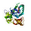
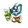
| Entry | Database: PDB / ID: 3wnd | ||||||
|---|---|---|---|---|---|---|---|
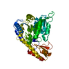
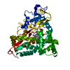
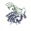
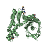
| Title | Crystal structure of EF-Pyl | ||||||
 Components Components | Protein translation elongation factor 1A | ||||||
 Keywords Keywords |  TRANSLATION / Structural Genomics / NPPSFA / National Project on Protein Structural and Functional Analyses / RIKEN Structural Genomics/Proteomics Initiative / RSGI / tRNA / elongation factor / pyrrolysine TRANSLATION / Structural Genomics / NPPSFA / National Project on Protein Structural and Functional Analyses / RIKEN Structural Genomics/Proteomics Initiative / RSGI / tRNA / elongation factor / pyrrolysine | ||||||
| Function / homology |  Function and homology information Function and homology information | ||||||
| Biological species |  Methanosarcina mazei (archaea) Methanosarcina mazei (archaea) | ||||||
| Method | X-RAY DIFFRACTION / SYNCHROTRON / MOLECULAR REPLACEMENT / Resolution: 1.55 Å | ||||||
 Authors Authors | Yanagisawa, T. / Ishii, R. / Fukunaga, R. / Sengoku, T. / Yokoyama, S. / RIKEN Structural Genomics/Proteomics Initiative (RSGI) | ||||||
 Citation Citation | Journal: J. Struct. Funct. Genomics / Year: 2015 Title: A SelB/EF-Tu/aIF2 gamma-like protein from Methanosarcina mazei in the GTP-bound form binds cysteinyl-tRNA(Cys.). Authors: Yanagisawa, T. / Ishii, R. / Hikida, Y. / Fukunaga, R. / Sengoku, T. / Sekine, S. / Yokoyama, S. | ||||||
| History |
|
- Structure visualization
| Structure viewer | Molecule: MolmilJmol/JSmol |
|---|
- Downloads & links
-Download
| PDBx/mmCIF format | 3wnd.cif.gz | 168.6 KB | Display | PDBx/mmCIF format |
|---|---|---|---|---|
| PDB format | pdb3wnd.ent.gz | 133.1 KB | Display | PDB format |
| PDBx/mmJSON format | 3wnd.json.gz | Tree view | PDBx/mmJSON format | |
| Others |  Other downloads Other downloads |
-Validation report
| Arichive directory | https://data.pdbj.org/pub/pdb/validation_reports/wn/3wndftp://data.pdbj.org/pub/pdb/validation_reports/wn/3wnd | HTTPS FTP |
|---|
-Related structure data
| Related structure data |  3wnbSC  3wncC S: Starting model for refinement C: citing same article ( |
|---|---|
| Similar structure data | |
| Other databases |


-Links
 PDBj
PDBj
- Assembly
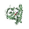
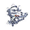
| Deposited unit | 
| |||||||||
|---|---|---|---|---|---|---|---|---|---|---|
| 1 |
| |||||||||
| Unit cell |
| |||||||||
| Components on special symmetry positions |
|
-Components
| #1: Protein | Mass: 40493.262 Da / Num. of mol.: 1 Source method: isolated from a genetically manipulated source Source: (gene. exp.) Methanosarcina mazei (archaea) / Strain: JCM9314 / Gene: MM_1309 / Plasmid: pET28c / Production host:  Escherichia coli (E. coli) / Strain (production host): BL21-Gold(DE3) / References: UniProt: Q8PXB3 Escherichia coli (E. coli) / Strain (production host): BL21-Gold(DE3) / References: UniProt: Q8PXB3 | ||
|---|---|---|---|
| #2: Chemical | Citric acid  Mass: 192.124 Da / Num. of mol.: 2 / Source method: obtained synthetically / Formula: C6H8O7 Mass: 192.124 Da / Num. of mol.: 2 / Source method: obtained synthetically / Formula: C6H8O7#3: Water | ChemComp-HOH / | Water Mass: 18.015 Da / Num. of mol.: 307 / Source method: isolated from a natural source / Formula: H2O Mass: 18.015 Da / Num. of mol.: 307 / Source method: isolated from a natural source / Formula: H2O |
-Experimental details
-Experiment
| Experiment | Method: X-RAY DIFFRACTION / Number of used crystals: 1 |
|---|
- Sample preparation
| Crystal | Density Matthews: 2.43 Å3/Da / Density % sol: 49.42 % |
|---|---|
| Crystal grow | Temperature: 277 K / Method: vapor diffusion, hanging drop / pH: 4.5 Details: Na/Acetate, Na/Citrate, pH 4.5, VAPOR DIFFUSION, HANGING DROP, temperature 277K |
-Data collection
| Diffraction | Mean temperature: 100 K |
|---|---|
| Diffraction source | Source: SYNCHROTRON / Site: SPring-8  / Beamline: BL41XU / Wavelength: 1 Å / Beamline: BL41XU / Wavelength: 1 Å |
| Detector | Type: RAYONIX MX225HE / Detector: CCD / Date: Apr 27, 2012 |
| Radiation | Monochromator: Rotated-inclined double-crystal monochromator, Si(111) Protocol: SINGLE WAVELENGTH / Monochromatic (M) / Laue (L): M / Scattering type: x-ray |
| Radiation wavelength | Wavelength: 1 Å / Relative weight: 1 |
| Reflection | Resolution: 1.55→50 Å / Num. obs: 57020 / % possible obs: 98.1 % / Redundancy: 4.8 % / Rsym value: 0.089 |
- Processing
| Software |
| ||||||||||||||||||||||||||||||||||||||||||||||||||||||||||||
|---|---|---|---|---|---|---|---|---|---|---|---|---|---|---|---|---|---|---|---|---|---|---|---|---|---|---|---|---|---|---|---|---|---|---|---|---|---|---|---|---|---|---|---|---|---|---|---|---|---|---|---|---|---|---|---|---|---|---|---|---|---|
| Refinement | Method to determine structure: MOLECULAR REPLACEMENT Starting model: 3WNB Resolution: 1.55→27.15 Å / Cor.coef. Fo:Fc: 0.963 / Cor.coef. Fo:Fc free: 0.954 / Occupancy max: 1 / Occupancy min: 0 / SU B: 3.19 / SU ML: 0.052 / Cross valid method: THROUGHOUT / ESU R: 0.104 / ESU R Free: 0.084 / Stereochemistry target values: MAXIMUM LIKELIHOOD Details: HYDROGENS HAVE BEEN USED IF PRESENT IN THE INPUT U VALUES: REFINED INDIVIDUALLY
| ||||||||||||||||||||||||||||||||||||||||||||||||||||||||||||
| Solvent computation | Ion probe radii: 0.8 Å / Shrinkage radii: 0.8 Å / VDW probe radii: 1.2 Å / Solvent model: MASK | ||||||||||||||||||||||||||||||||||||||||||||||||||||||||||||
| Displacement parameters | Biso max: 107.64 Å2 / Biso mean: 24.0502 Å2 / Biso min: 6.62 Å2
| ||||||||||||||||||||||||||||||||||||||||||||||||||||||||||||
| Refinement step | Cycle: LAST / Resolution: 1.55→27.15 Å
| ||||||||||||||||||||||||||||||||||||||||||||||||||||||||||||
| Refine LS restraints |
| ||||||||||||||||||||||||||||||||||||||||||||||||||||||||||||
| LS refinement shell | Resolution: 1.55→1.59 Å / Total num. of bins used: 20
|
