 Movie
Movie Controller

[English] 日本語
 Yorodumi
- PDB-3wmu: The structure of an anti-cancer lectin mytilec apo-form from the ... -
+ Open data

- Basic information
| Entry | Database: PDB / ID: 3wmu | ||||||
|---|---|---|---|---|---|---|---|
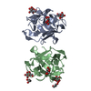
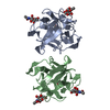
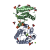
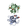
| Title | The structure of an anti-cancer lectin mytilec apo-form from the mussel Mytilus galloprovincialis | ||||||
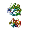
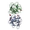
 Components Components | Lectin | ||||||
 Keywords Keywords | SUGAR BINDING PROTEIN / LECTIN / CARBOHYDRATE | ||||||
| Function / homology | galactose binding / Trefoil (Acidic Fibroblast Growth Factor, subunit A) - #50 / Trefoil (Acidic Fibroblast Growth Factor, subunit A) / Trefoil / killing of cells of another organism / Mainly Beta / Alpha-D-galactose-binding lectin Function and homology information Function and homology information | ||||||
| Biological species |  Mytilus galloprovincialis (Mediterranean mussel) Mytilus galloprovincialis (Mediterranean mussel) | ||||||
| Method | X-RAY DIFFRACTION / SYNCHROTRON / MOLECULAR REPLACEMENT / Resolution: 1.1 Å | ||||||
 Authors Authors | Terada, D. / Kawai, F. / Noguchi, H. / Unzai, S. / Park, S.-Y. / Ozeki, Y. / Tame, J.R.H. | ||||||
 Citation Citation | Journal: Sci Rep / Year: 2016 Title: Crystal structure of MytiLec, a galactose-binding lectin from the mussel Mytilus galloprovincialis with cytotoxicity against certain cancer cell types Authors: Terada, D. / Kawai, F. / Noguchi, H. / Unzai, S. / Hasan, I. / Fujii, Y. / Park, S.-Y. / Ozeki, Y. / Tame, J.R.H. | ||||||
| History |
|
- Structure visualization
| Structure viewer | Molecule: MolmilJmol/JSmol |
|---|
- Downloads & links
-Download
| PDBx/mmCIF format | 3wmu.cif.gz | 146.2 KB | Display | PDBx/mmCIF format |
|---|---|---|---|---|
| PDB format | pdb3wmu.ent.gz | 115.6 KB | Display | PDB format |
| PDBx/mmJSON format | 3wmu.json.gz | Tree view | PDBx/mmJSON format | |
| Others |  Other downloads Other downloads |
-Validation report
| Arichive directory | https://data.pdbj.org/pub/pdb/validation_reports/wm/3wmuftp://data.pdbj.org/pub/pdb/validation_reports/wm/3wmu | HTTPS FTP |
|---|
-Related structure data


-Links
 PDBj
PDBj- Assembly
| Deposited unit | 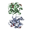
| ||||||||
|---|---|---|---|---|---|---|---|---|---|
| 1 |
| ||||||||
| Unit cell |
|
-Components
| #1: Protein | / MytiLec Mass: 17195.615 Da / Num. of mol.: 2 / Mutation: T1A Source method: isolated from a genetically manipulated source Source: (gene. exp.) Mytilus galloprovincialis (Mediterranean mussel)Production host:  Escherichia coli (E. coli) / References: UniProt: B3EWR1 Escherichia coli (E. coli) / References: UniProt: B3EWR1#2: Chemical | ChemComp-GOL / Glycerol  Mass: 92.094 Da / Num. of mol.: 6 / Source method: obtained synthetically / Formula: C3H8O3 Mass: 92.094 Da / Num. of mol.: 6 / Source method: obtained synthetically / Formula: C3H8O3#3: Water | ChemComp-HOH / | Water Mass: 18.015 Da / Num. of mol.: 341 / Source method: isolated from a natural source / Formula: H2O Mass: 18.015 Da / Num. of mol.: 341 / Source method: isolated from a natural source / Formula: H2O |
|---|
-Experimental details
-Experiment
| Experiment | Method: X-RAY DIFFRACTION / Number of used crystals: 1 |
|---|
- Sample preparation
| Crystal | Density Matthews: 2.27 Å3/Da / Density % sol: 45.92 % |
|---|---|
| Crystal grow | Temperature: 293 K / Method: vapor diffusion, hanging drop / pH: 7.5 Details: Sodiumu acetate, HEPES, Glycerol, PEG4000, pH 7.5, VAPOR DIFFUSION, HANGING DROP, temperature 293K |
-Data collection
| Diffraction | Mean temperature: 95 K |
|---|---|
| Diffraction source | Source: SYNCHROTRON / Site: Photon Factory  / Beamline: BL-1A / Wavelength: 1.1 Å / Beamline: BL-1A / Wavelength: 1.1 Å |
| Detector | Type: DECTRIS PILATUS 2M-F / Detector: PIXEL / Date: May 24, 2013 |
| Radiation | Monochromator: Si(111) / Protocol: SINGLE WAVELENGTH / Monochromatic (M) / Laue (L): M / Scattering type: x-ray |
| Radiation wavelength | Wavelength: 1.1 Å / Relative weight: 1 |
| Reflection | Resolution: 1.1→50 Å / Num. obs: 120340 / % possible obs: 96 % / Observed criterion σ(F): 0 / Observed criterion σ(I): 0 |
| Reflection shell | Resolution: 1.1→1.12 Å / Rmerge(I) obs: 0.298 / % possible all: 89.6 |
- Processing
| Software |
| |||||||||||||||||||||||||||||||||||||||||||||||||||||||||||||||||||||||||||
|---|---|---|---|---|---|---|---|---|---|---|---|---|---|---|---|---|---|---|---|---|---|---|---|---|---|---|---|---|---|---|---|---|---|---|---|---|---|---|---|---|---|---|---|---|---|---|---|---|---|---|---|---|---|---|---|---|---|---|---|---|---|---|---|---|---|---|---|---|---|---|---|---|---|---|---|---|
| Refinement | Method to determine structure: MOLECULAR REPLACEMENT / Resolution: 1.1→27.48 Å / Cor.coef. Fo:Fc: 0.974 / Cor.coef. Fo:Fc free: 0.967 / SU B: 0.857 / SU ML: 0.019 / Cross valid method: THROUGHOUT / ESU R: 0.028 / ESU R Free: 0.029 / Stereochemistry target values: MAXIMUM LIKELIHOOD / Details: HYDROGENS HAVE BEEN ADDED IN THE RIDING POSITIONS
| |||||||||||||||||||||||||||||||||||||||||||||||||||||||||||||||||||||||||||
| Solvent computation | Ion probe radii: 0.8 Å / Shrinkage radii: 0.8 Å / VDW probe radii: 1.2 Å / Solvent model: MASK | |||||||||||||||||||||||||||||||||||||||||||||||||||||||||||||||||||||||||||
| Displacement parameters | Biso mean: 11.114 Å2
| |||||||||||||||||||||||||||||||||||||||||||||||||||||||||||||||||||||||||||
| Refinement step | Cycle: LAST / Resolution: 1.1→27.48 Å
| |||||||||||||||||||||||||||||||||||||||||||||||||||||||||||||||||||||||||||
| Refine LS restraints |
| |||||||||||||||||||||||||||||||||||||||||||||||||||||||||||||||||||||||||||
| LS refinement shell | Resolution: 1.1→1.129 Å / Total num. of bins used: 20
|
