 Movie
Movie Controller

[English] 日本語
 Yorodumi
- PDB-3wi5: Crystal structure of the Loop 7 mutant PorB from Neisseria mening... -
+ Open data

- Basic information
| Entry | Database: PDB / ID: 3wi5 | ||||||
|---|---|---|---|---|---|---|---|
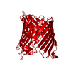
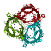
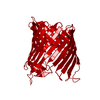
| Title | Crystal structure of the Loop 7 mutant PorB from Neisseria meningitidis serogroup B | ||||||
 Components Components | Major outer membrane protein P.IB | ||||||
 Keywords Keywords |  MEMBRANE PROTEIN / beta-barrel / porin / outer membrane MEMBRANE PROTEIN / beta-barrel / porin / outer membrane | ||||||
| Function / homology |  Function and homology information Function and homology informationToll Like Receptor TLR1:TLR2 Cascade / MyD88 deficiency (TLR2/4) / IRAK4 deficiency (TLR2/4) / MyD88:MAL(TIRAP) cascade initiated on plasma membrane / porin activity / pore complex / monoatomic ion transmembrane transport / cell outer membrane / ER-Phagosome pathwaySimilarity search - Function | ||||||
| Biological species |  Neisseria meningitidis (bacteria) Neisseria meningitidis (bacteria) | ||||||
| Method | X-RAY DIFFRACTION / SYNCHROTRON / MOLECULAR REPLACEMENT / Resolution: 2.4 Å | ||||||
 Authors Authors | Kattner, C. / Toussi, D. / Wetzler, L.M. / Ruppel, N. / Massari, P. / Tanabe, M. | ||||||
 Citation Citation | Journal: J.Struct.Biol. / Year: 2014 Title: Crystallographic analysis of Neisseria meningitidis PorB extracellular loops potentially implicated in TLR2 recognition. Authors: Kattner, C. / Toussi, D.N. / Zaucha, J. / Wetzler, L.M. / Ruppel, N. / Zachariae, U. / Massari, P. / Tanabe, M. | ||||||
| History |
|
- Structure visualization
| Structure viewer | Molecule: MolmilJmol/JSmol |
|---|
- Downloads & links
-Download
| PDBx/mmCIF format | 3wi5.cif.gz | 74.7 KB | Display | PDBx/mmCIF format |
|---|---|---|---|---|
| PDB format | pdb3wi5.ent.gz | 55.8 KB | Display | PDB format |
| PDBx/mmJSON format | 3wi5.json.gz | Tree view | PDBx/mmJSON format | |
| Others |  Other downloads Other downloads |
-Validation report
| Arichive directory | https://data.pdbj.org/pub/pdb/validation_reports/wi/3wi5ftp://data.pdbj.org/pub/pdb/validation_reports/wi/3wi5 | HTTPS FTP |
|---|
-Related structure data
| Related structure data | 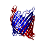 3wi4C  3vztS C: citing same article ( S: Starting model for refinement |
|---|---|
| Similar structure data |


-Links
 PDBj
PDBj


- Assembly
| Deposited unit | 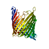
| ||||||||
|---|---|---|---|---|---|---|---|---|---|
| 1 | 
| ||||||||
| Unit cell |
|
-Components
| #1: Protein | Mass: 33825.363 Da / Num. of mol.: 1 / Mutation: D259A, D260K, E266R Source method: isolated from a genetically manipulated source Source: (gene. exp.) Neisseria meningitidis (bacteria) / Strain: MC58 / Gene: porB, NMB2039 / Production host: Escherichia coli (E. coli) / References: UniProt: P30690 | ||
|---|---|---|---|
| #2: Chemical | Citric acid  Mass: 189.100 Da / Num. of mol.: 2 / Source method: obtained synthetically / Formula: C6H5O7 Mass: 189.100 Da / Num. of mol.: 2 / Source method: obtained synthetically / Formula: C6H5O7#3: Water | ChemComp-HOH / | Water Mass: 18.015 Da / Num. of mol.: 82 / Source method: isolated from a natural source / Formula: H2O Mass: 18.015 Da / Num. of mol.: 82 / Source method: isolated from a natural source / Formula: H2O |
-Experimental details
-Experiment
| Experiment | Method: X-RAY DIFFRACTION / Number of used crystals: 1 |
|---|
- Sample preparation
| Crystal grow | Temperature: 293 K / Method: vapor diffusion, hanging drop / pH: 7.5 Details: 65mM HEPES pH 7.5, 1.1M tri-sodium citrate, VAPOR DIFFUSION, HANGING DROP, temperature 293K |
|---|
-Data collection
| Diffraction | Mean temperature: 100 K |
|---|---|
| Diffraction source | Source: SYNCHROTRON / Site: SLS  / Beamline: X06DA / Wavelength: 1 Å / Beamline: X06DA / Wavelength: 1 Å |
| Detector | Type: DECTRIS PILATUS 2M / Detector: PIXEL / Date: Nov 22, 2012 |
| Radiation | Monochromator: toroidal mirror / Protocol: SINGLE WAVELENGTH / Monochromatic (M) / Laue (L): M / Scattering type: x-ray |
| Radiation wavelength | Wavelength: 1 Å / Relative weight: 1 |
| Reflection | Resolution: 2.4→50 Å / Num. obs: 25466 / % possible obs: 92.2 % / Observed criterion σ(F): 17.4 / Observed criterion σ(I): 1.55 |
| Reflection shell | Resolution: 2.4→2.45 Å / % possible all: 73 |
- Processing
| Software |
| |||||||||||||||||||||||||||||||||||||||||||||
|---|---|---|---|---|---|---|---|---|---|---|---|---|---|---|---|---|---|---|---|---|---|---|---|---|---|---|---|---|---|---|---|---|---|---|---|---|---|---|---|---|---|---|---|---|---|---|
| Refinement | Method to determine structure: MOLECULAR REPLACEMENT Starting model: 3VZT Resolution: 2.4→50 Å / Cor.coef. Fo:Fc: 0.932 / Cor.coef. Fo:Fc free: 0.916 / Cross valid method: THROUGHOUT / ESU R: 0.246 / ESU R Free: 0.211 / Stereochemistry target values: MAXIMUM LIKELIHOOD / Details: HYDROGENS HAVE BEEN USED IF PRESENT IN THE INPUT
| |||||||||||||||||||||||||||||||||||||||||||||
| Solvent computation | Ion probe radii: 0.8 Å / Shrinkage radii: 0.8 Å / VDW probe radii: 1.2 Å / Solvent model: MASK | |||||||||||||||||||||||||||||||||||||||||||||
| Displacement parameters | Biso mean: 51.113 Å2
| |||||||||||||||||||||||||||||||||||||||||||||
| Refinement step | Cycle: LAST / Resolution: 2.4→50 Å
| |||||||||||||||||||||||||||||||||||||||||||||
| Refine LS restraints |
| |||||||||||||||||||||||||||||||||||||||||||||
| LS refinement shell | Resolution: 2.404→2.466 Å / Total num. of bins used: 20
|
