 Movie
Movie Controller


+ Open data

- Basic information
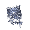
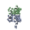
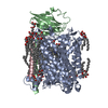
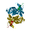
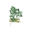
| Entry | Database: PDB / ID: 3wbm | ||||||
|---|---|---|---|---|---|---|---|
| Title | Crystal Structure of protein-RNA complex | ||||||
 Components Components |
| ||||||
 Keywords Keywords | RNA BINDING PROTEIN/RNA / PROTEIN-RNA COMPLEX / DNA/RNA BINDING / RNA BINDING PROTEIN-RNA complex | ||||||
| Function / homology |  Function and homology information Function and homology informationchromosome condensation /  chromosome / double-stranded DNA binding / Hydrolases; Acting on ester bonds / RNA binding / cytoplasm chromosome / double-stranded DNA binding / Hydrolases; Acting on ester bonds / RNA binding / cytoplasmSimilarity search - Function | ||||||
| Biological species |   Sulfolobus shibatae (archaea) Sulfolobus shibatae (archaea)synthetic construct (others) | ||||||
| Method | X-RAY DIFFRACTION / SYNCHROTRON / MOLECULAR REPLACEMENT / Resolution: 2 Å | ||||||
 Authors Authors | Ding, J. / Wang, D.C. | ||||||
 Citation Citation | Journal: J.Biol.Chem. / Year: 2014 Title: Biochemical and structural insights into RNA binding by Ssh10b, a member of the highly conserved Sac10b protein family in Archaea. Authors: Guo, L. / Ding, J. / Guo, R. / Hou, Y. / Wang, D.C. / Huang, L. | ||||||
| History |
|
- Structure visualization
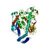
| Structure viewer | Molecule: MolmilJmol/JSmol |
|---|
- Downloads & links
-Download
| PDBx/mmCIF format | 3wbm.cif.gz | 113.6 KB | Display | PDBx/mmCIF format |
|---|---|---|---|---|
| PDB format | pdb3wbm.ent.gz | 84.4 KB | Display | PDB format |
| PDBx/mmJSON format | 3wbm.json.gz | Tree view | PDBx/mmJSON format | |
| Others |  Other downloads Other downloads |
-Validation report
| Arichive directory | https://data.pdbj.org/pub/pdb/validation_reports/wb/3wbmftp://data.pdbj.org/pub/pdb/validation_reports/wb/3wbm | HTTPS FTP |
|---|
-Related structure data
| Related structure data | 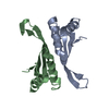 1h0xS S: Starting model for refinement |
|---|---|
| Similar structure data |


-Links
 PDBj
PDBj


- Assembly
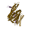
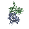
| Deposited unit | 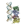
| ||||||||
|---|---|---|---|---|---|---|---|---|---|
| 1 |
| ||||||||
| Unit cell |
| ||||||||
| Components on special symmetry positions |
|

-Components
| #1: Protein | Mass: 10601.442 Da / Num. of mol.: 4 Source method: isolated from a genetically manipulated source Source: (gene. exp.) Sulfolobus shibatae (archaea) / Strain: 2286 / Gene: albA1, ssh10b / Plasmid: pET30a / Production host:  Escherichia coli BL21(DE3) (bacteria) / References: UniProt: P60848 Escherichia coli BL21(DE3) (bacteria) / References: UniProt: P60848#2: RNA chain | Mass: 7949.771 Da / Num. of mol.: 2 / Source method: obtained synthetically / Source: (synth.) synthetic construct (others) #3: Water | ChemComp-HOH / | Water Mass: 18.015 Da / Num. of mol.: 310 / Source method: isolated from a natural source / Formula: H2O Mass: 18.015 Da / Num. of mol.: 310 / Source method: isolated from a natural source / Formula: H2O |
|---|
-Experimental details
-Experiment
| Experiment | Method: X-RAY DIFFRACTION / Number of used crystals: 1 |
|---|
- Sample preparation
| Crystal | Density Matthews: 2.43 Å3/Da / Density % sol: 49.4 % |
|---|---|
| Crystal grow | Temperature: 293 K / Method: vapor diffusion, hanging drop / pH: 7 Details: 18% (v/v) PEG 5000 MME, 100mM Bis-Tris, 200mM sodium malonate, pH 7.0, VAPOR DIFFUSION, HANGING DROP, temperature 293K |
-Data collection
| Diffraction | Mean temperature: 95 K |
|---|---|
| Diffraction source | Source: SYNCHROTRON / Site: Photon Factory  / Beamline: BL-17A / Wavelength: 0.9642 Å / Beamline: BL-17A / Wavelength: 0.9642 Å |
| Detector | Type: ADSC QUANTUM 315r / Detector: CCD / Date: Jun 6, 2012 |
| Radiation | Protocol: SINGLE WAVELENGTH / Monochromatic (M) / Laue (L): M / Scattering type: x-ray |
| Radiation wavelength | Wavelength: 0.9642 Å / Relative weight: 1 |
| Reflection | Resolution: 2→47.84 Å / Num. all: 37892 / Num. obs: 37589 / % possible obs: 99.2 % / Observed criterion σ(F): 0 / Observed criterion σ(I): 0 / Redundancy: 3.6 % / Biso Wilson estimate: 18.85 Å2 / Rmerge(I) obs: 0.074 / Rsym value: 0.074 / Net I/σ(I): 11 |
| Reflection shell | Resolution: 2→2.11 Å / Redundancy: 3.6 % / Rmerge(I) obs: 0.407 / Mean I/σ(I) obs: 3.1 / Num. unique all: 5444 / Rsym value: 0.407 / % possible all: 98.7 |
- Processing
| Software |
| ||||||||||||||||||||||||||||||||||||||||||||||||||||||||||||||||||||||||||||||||||||
|---|---|---|---|---|---|---|---|---|---|---|---|---|---|---|---|---|---|---|---|---|---|---|---|---|---|---|---|---|---|---|---|---|---|---|---|---|---|---|---|---|---|---|---|---|---|---|---|---|---|---|---|---|---|---|---|---|---|---|---|---|---|---|---|---|---|---|---|---|---|---|---|---|---|---|---|---|---|---|---|---|---|---|---|---|---|
| Refinement | Method to determine structure: MOLECULAR REPLACEMENT Starting model: PDB ENTRY 1H0X Resolution: 2→43.804 Å / Occupancy max: 1 / Occupancy min: 1 / FOM work R set: 0.8165 / SU ML: 0.23 / σ(F): 1.35 / Phase error: 25.69 / Stereochemistry target values: ML
| ||||||||||||||||||||||||||||||||||||||||||||||||||||||||||||||||||||||||||||||||||||
| Solvent computation | Shrinkage radii: 0.98 Å / VDW probe radii: 1.2 Å / Solvent model: FLAT BULK SOLVENT MODEL / Bsol: 36.773 Å2 / ksol: 0.351 e/Å3 | ||||||||||||||||||||||||||||||||||||||||||||||||||||||||||||||||||||||||||||||||||||
| Displacement parameters | Biso max: 110.96 Å2 / Biso mean: 40.5243 Å2 / Biso min: 14.49 Å2
| ||||||||||||||||||||||||||||||||||||||||||||||||||||||||||||||||||||||||||||||||||||
| Refine analyze | Luzzati coordinate error obs: 0.23 Å | ||||||||||||||||||||||||||||||||||||||||||||||||||||||||||||||||||||||||||||||||||||
| Refinement step | Cycle: LAST / Resolution: 2→43.804 Å
| ||||||||||||||||||||||||||||||||||||||||||||||||||||||||||||||||||||||||||||||||||||
| Refine LS restraints |
| ||||||||||||||||||||||||||||||||||||||||||||||||||||||||||||||||||||||||||||||||||||
| LS refinement shell | Refine-ID: X-RAY DIFFRACTION / Total num. of bins used: 13
|
