 Movie
Movie Controller

[English] 日本語
 Yorodumi
- PDB-3wbf: Crystal Structure of meso-diaminopimelate dehydrogenase from Symb... -
+ Open data

- Basic information
| Entry | Database: PDB / ID: 3wbf | |||||||||
|---|---|---|---|---|---|---|---|---|---|---|
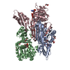
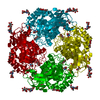
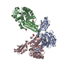
| Title | Crystal Structure of meso-diaminopimelate dehydrogenase from Symbiobacterium thermophilum co-crystallized with NADP+ and DAP | |||||||||
 Components Components | Diaminopimelate dehydrogenase | |||||||||
 Keywords Keywords | OXIDOREDUCTASE / domain motion / thermo-stable / d-amino acid dehydrogenase | |||||||||
| Function / homology |  Function and homology informationdiaminopimelate dehydrogenase / diaminopimelate dehydrogenase activity / diaminopimelate biosynthetic process / lysine biosynthetic process via diaminopimelate / nucleotide binding Function and homology informationdiaminopimelate dehydrogenase / diaminopimelate dehydrogenase activity / diaminopimelate biosynthetic process / lysine biosynthetic process via diaminopimelate / nucleotide bindingSimilarity search - Function | |||||||||
| Biological species |  Symbiobacterium thermophilum (bacteria) Symbiobacterium thermophilum (bacteria) | |||||||||
| Method | X-RAY DIFFRACTION / SYNCHROTRON / MOLECULAR REPLACEMENT / molecular replacement / Resolution: 2.12 Å | |||||||||
 Authors Authors | Liu, W.D. / Li, Z. / Huang, C.H. / Guo, R.T. / Wu, Q.Q. / Zhu, D.M. | |||||||||
 Citation Citation | Journal: Chembiochem / Year: 2014 Title: Structural and mutational studies on the unusual substrate specificity of meso-diaminopimelate dehydrogenase from Symbiobacterium thermophilum. Authors: Liu, W. / Li, Z. / Huang, C.H. / Guo, R.T. / Zhao, L. / Zhang, D. / Chen, X. / Wu, Q. / Zhu, D. | |||||||||
| History |
|
- Structure visualization
| Structure viewer | Molecule: MolmilJmol/JSmol |
|---|
- Downloads & links
-Download
| PDBx/mmCIF format | 3wbf.cif.gz | 221 KB | Display | PDBx/mmCIF format |
|---|---|---|---|---|
| PDB format | pdb3wbf.ent.gz | 174.1 KB | Display | PDB format |
| PDBx/mmJSON format | 3wbf.json.gz | Tree view | PDBx/mmJSON format | |
| Others |  Other downloads Other downloads |
-Validation report
| Arichive directory | https://data.pdbj.org/pub/pdb/validation_reports/wb/3wbfftp://data.pdbj.org/pub/pdb/validation_reports/wb/3wbf | HTTPS FTP |
|---|
-Related structure data
| Related structure data |  3wb9C  3wbbC 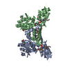 3bioS C: citing same article ( S: Starting model for refinement |
|---|---|
| Similar structure data |
-Links
 PDBj
PDBj- Assembly
| Deposited unit | 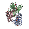
| ||||||||
|---|---|---|---|---|---|---|---|---|---|
| 1 | 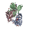
| ||||||||
| Unit cell |
|
-Components
| #1: Protein | Mass: 33312.410 Da / Num. of mol.: 3 Source method: isolated from a genetically manipulated source Source: (gene. exp.) Symbiobacterium thermophilum (bacteria)Strain: T / IAM 14863 / Gene: meso-dapdh, STH1425 / Plasmid: pET32 / Production host: Escherichia coli (E. coli) / Strain (production host): BL21(DE3) / References: UniProt: Q67PI3, diaminopimelate dehydrogenase#2: Chemical | Diaminopimelic acid  Type: L-peptide linking / Mass: 190.197 Da / Num. of mol.: 3 / Source method: obtained synthetically / Formula: C7H14N2O4 Type: L-peptide linking / Mass: 190.197 Da / Num. of mol.: 3 / Source method: obtained synthetically / Formula: C7H14N2O4#3: Chemical | ChemComp-GOL / Glycerol  Mass: 92.094 Da / Num. of mol.: 16 / Source method: obtained synthetically / Formula: C3H8O3 Mass: 92.094 Da / Num. of mol.: 16 / Source method: obtained synthetically / Formula: C3H8O3#4: Chemical | Nicotinamide adenine dinucleotide phosphate  Mass: 743.405 Da / Num. of mol.: 3 / Source method: obtained synthetically / Formula: C21H28N7O17P3 Mass: 743.405 Da / Num. of mol.: 3 / Source method: obtained synthetically / Formula: C21H28N7O17P3#5: Water | ChemComp-HOH / | Water Mass: 18.015 Da / Num. of mol.: 1406 / Source method: isolated from a natural source / Formula: H2O Mass: 18.015 Da / Num. of mol.: 1406 / Source method: isolated from a natural source / Formula: H2O |
|---|
-Experimental details
-Experiment
| Experiment | Method: X-RAY DIFFRACTION / Number of used crystals: 1 |
|---|
- Sample preparation
| Crystal | Density Matthews: 2.74 Å3/Da / Density % sol: 55.07 % / Mosaicity: 0.932 ° |
|---|---|
| Crystal grow | Temperature: 288 K / Method: vapor diffusion, sitting drop / pH: 7.5 Details: MPD, PEG 6000, NADP+, DAP,, pH 7.5, vapor diffusion, sitting drop, temperature 288K |
-Data collection
| Diffraction | Mean temperature: 100 K | |||||||||||||||||||||||||||||||||||||||||||||||||||||||||||||||||||||||||||||
|---|---|---|---|---|---|---|---|---|---|---|---|---|---|---|---|---|---|---|---|---|---|---|---|---|---|---|---|---|---|---|---|---|---|---|---|---|---|---|---|---|---|---|---|---|---|---|---|---|---|---|---|---|---|---|---|---|---|---|---|---|---|---|---|---|---|---|---|---|---|---|---|---|---|---|---|---|---|---|
| Diffraction source | Source: SYNCHROTRON / Site: NSRRC  / Beamline: BL13B1 / Wavelength: 1 Å / Beamline: BL13B1 / Wavelength: 1 Å | |||||||||||||||||||||||||||||||||||||||||||||||||||||||||||||||||||||||||||||
| Detector | Type: ADSC QUANTUM 315 / Detector: CCD / Date: Dec 25, 2012 | |||||||||||||||||||||||||||||||||||||||||||||||||||||||||||||||||||||||||||||
| Radiation | Protocol: SINGLE WAVELENGTH / Monochromatic (M) / Laue (L): M / Scattering type: x-ray | |||||||||||||||||||||||||||||||||||||||||||||||||||||||||||||||||||||||||||||
| Radiation wavelength | Wavelength: 1 Å / Relative weight: 1 | |||||||||||||||||||||||||||||||||||||||||||||||||||||||||||||||||||||||||||||
| Reflection | Resolution: 2.12→50 Å / Num. all: 61605 / Num. obs: 61060 / % possible obs: 99.2 % / Redundancy: 3.6 % / Rmerge(I) obs: 0.099 / Χ2: 1.324 / Net I/σ(I): 7.1 | |||||||||||||||||||||||||||||||||||||||||||||||||||||||||||||||||||||||||||||
| Reflection shell |
|
-Phasing
| Phasing | Method: molecular replacement |
|---|
- Processing
| Software |
| |||||||||||||||||||||||||
|---|---|---|---|---|---|---|---|---|---|---|---|---|---|---|---|---|---|---|---|---|---|---|---|---|---|---|
| Refinement | Method to determine structure: MOLECULAR REPLACEMENT Starting model: 3BIO Resolution: 2.12→30 Å / Occupancy max: 1 / Occupancy min: 0.5 / FOM work R set: 0.8134 / σ(F): 0 / Stereochemistry target values: Engh & Huber
| |||||||||||||||||||||||||
| Solvent computation | Bsol: 60.4694 Å2 | |||||||||||||||||||||||||
| Displacement parameters | Biso max: 88.05 Å2 / Biso mean: 29.0927 Å2 / Biso min: 8.62 Å2
| |||||||||||||||||||||||||
| Refinement step | Cycle: LAST / Resolution: 2.12→30 Å
| |||||||||||||||||||||||||
| Refine LS restraints |
| |||||||||||||||||||||||||
| Xplor file |
|
