 Movie
Movie Controller


+ Open data

- Basic information
| Entry | Database: PDB / ID: 3w10 | ||||||
|---|---|---|---|---|---|---|---|
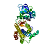
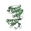
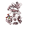
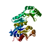
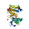
| Title | Aurora kinase A complexed to pyrazole aminoquinoline I | ||||||
 Components Components | Aurora kinase A | ||||||
 Keywords Keywords | TRANSFERASE/TRANSFERASE INHIBITOR / kinase / pyrazole aminoquinoline / TRANSFERASE-TRANSFERASE INHIBITOR complex | ||||||
| Function / homology |  Function and homology information Function and homology informationInteraction between PHLDA1 and AURKA / regulation of centrosome cycle / axon hillock / spindle assembly involved in female meiosis I / cilium disassembly / spindle pole centrosome / positive regulation of oocyte maturation / histone H3S10 kinase activity / chromosome passenger complex / pronucleus ...Interaction between PHLDA1 and AURKA / regulation of centrosome cycle / axon hillock / spindle assembly involved in female meiosis I / cilium disassembly / spindle pole centrosome / positive regulation of oocyte maturation / histone H3S10 kinase activity / chromosome passenger complex / pronucleus / meiotic spindle / mitotic centrosome separation / germinal vesicle / protein localization to centrosome / anterior/posterior axis specification / centrosome localization / neuron projection extension / positive regulation of mitochondrial fission / spindle organization / mitotic spindle pole / SUMOylation of DNA replication proteins / spindle midzone / regulation of G2/M transition of mitotic cell cycle / centriole / protein serine/threonine/tyrosine kinase activity / positive regulation of mitotic cell cycle / AURKA Activation by TPX2 / mitotic spindle organization / positive regulation of mitotic nuclear division / TP53 Regulates Transcription of Genes Involved in G2 Cell Cycle Arrest / ciliary basal body / negative regulation of protein binding / regulation of cytokinesis / regulation of signal transduction by p53 class mediator / molecular function activator activity / liver regeneration / FBXL7 down-regulates AURKA during mitotic entry and in early mitosis / regulation of protein stability / APC/C:Cdh1 mediated degradation of Cdc20 and other APC/C:Cdh1 targeted proteins in late mitosis/early G1 / mitotic spindle / kinetochore / spindle / response to wounding / microtubule cytoskeleton / G2/M transition of mitotic cell cycle / Regulation of PLK1 Activity at G2/M Transition / positive regulation of proteasomal ubiquitin-dependent protein catabolic process / mitotic cell cycle / midbody / proteasome-mediated ubiquitin-dependent protein catabolic process / basolateral plasma membrane / peptidyl-serine phosphorylation / Regulation of TP53 Activity through Phosphorylation / microtubule / postsynaptic density / protein autophosphorylation / non-specific serine/threonine protein kinase / protein kinase activity / protein heterodimerization activity / cell division / protein phosphorylation / negative regulation of gene expression / protein serine kinase activity / protein serine/threonine kinase activity / centrosome / glutamatergic synapse / apoptotic process / ubiquitin protein ligase binding / negative regulation of apoptotic process / protein kinase binding / perinuclear region of cytoplasm / nucleoplasm / ATP binding / nucleus / cytosolSimilarity search - Function | ||||||
| Biological species |  Homo sapiens (human) Homo sapiens (human) | ||||||
| Method | X-RAY DIFFRACTION / SYNCHROTRON / MOLECULAR REPLACEMENT / Resolution: 2.7 Å | ||||||
 Authors Authors | Oliveira, T.M. / Kairies, N.A. / Engh, R.A. | ||||||
 Citation Citation | Journal: To be Published Title: Flexibility and multiple conformations of the activation and glycine rich loops of Aurora A accompanying inhibitor binding Authors: Oliveira, T.M. / Kairies, N.A. / Engh, R.A. | ||||||
| History |
|
- Structure visualization
| Structure viewer | Molecule: MolmilJmol/JSmol |
|---|
- Downloads & links
-Download
| PDBx/mmCIF format | 3w10.cif.gz | 64 KB | Display | PDBx/mmCIF format |
|---|---|---|---|---|
| PDB format | pdb3w10.ent.gz | 50 KB | Display | PDB format |
| PDBx/mmJSON format | 3w10.json.gz | Tree view | PDBx/mmJSON format | |
| Others |  Other downloads Other downloads |
-Validation report
| Arichive directory | https://data.pdbj.org/pub/pdb/validation_reports/w1/3w10ftp://data.pdbj.org/pub/pdb/validation_reports/w1/3w10 | HTTPS FTP |
|---|
-Related structure data


-Links
 PDBj
PDBj


- Assembly
| Deposited unit | 
| ||||||||
|---|---|---|---|---|---|---|---|---|---|
| 1 |
| ||||||||
| Unit cell |
|
-Components
| #1: Protein | / Aurora 2 / Aurora/IPL1-related kinase 1 / ARK-1 / Aurora-related kinase 1 / hARK1 / Breast tumor- ...Aurora 2 / Aurora/IPL1-related kinase 1 / ARK-1 / Aurora-related kinase 1 / hARK1 / Breast tumor-amplified kinase / Serine/threonine-protein kinase 15 / Serine/threonine-protein kinase 6 / Serine/threonine-protein kinase aurora-A Mass: 32154.855 Da / Num. of mol.: 1 / Fragment: Domain, UNP RESIDUES 126-403 / Mutation: T287A, T288A Source method: isolated from a genetically manipulated source Source: (gene. exp.) Homo sapiens (human)Gene: AURKA, AIK, AIRK1, ARK1, AURA, AYK1, BTAK, IAK1, STK15, STK6 Production host:  Escherichia coli (E. coli) Escherichia coli (E. coli)References: UniProt: O14965, non-specific serine/threonine protein kinase |
|---|---|
| #2: Chemical | ChemComp-RO9 /   Mass: 330.383 Da / Num. of mol.: 1 / Source method: obtained synthetically / Formula: C20H18N4O Mass: 330.383 Da / Num. of mol.: 1 / Source method: obtained synthetically / Formula: C20H18N4O |
| #3: Water | ChemComp-HOH / Water Mass: 18.015 Da / Num. of mol.: 51 / Source method: isolated from a natural source / Formula: H2O Mass: 18.015 Da / Num. of mol.: 51 / Source method: isolated from a natural source / Formula: H2O |
-Experimental details
-Experiment
| Experiment | Method: X-RAY DIFFRACTION / Number of used crystals: 1 |
|---|
- Sample preparation
| Crystal | Density Matthews: 2.98 Å3/Da / Density % sol: 58.78 % |
|---|---|
| Crystal grow | Temperature: 298 K / Method: vapor diffusion, hanging drop / pH: 7 Details: PEG 3350, pH 7.0, VAPOR DIFFUSION, HANGING DROP, temperature 298K |
-Data collection
| Diffraction | Mean temperature: 298 K |
|---|---|
| Diffraction source | Source: SYNCHROTRON / Site: BESSY  / Beamline: 14.1 / Wavelength: 1.488 Å / Beamline: 14.1 / Wavelength: 1.488 Å |
| Detector | Type: MAR CCD 165 mm / Detector: CCD / Date: Oct 1, 2010 |
| Radiation | Monochromator: graphite / Protocol: SINGLE WAVELENGTH / Monochromatic (M) / Laue (L): M / Scattering type: x-ray |
| Radiation wavelength | Wavelength: 1.488 Å / Relative weight: 1 |
| Reflection | Resolution: 2.7→26.54 Å / Num. all: 11390 / Num. obs: 10633 / % possible obs: 70 % / Observed criterion σ(F): 3 / Observed criterion σ(I): 2 |
| Reflection shell | Resolution: 2.7→3 Å / % possible all: 70 |
- Processing
| Software |
| |||||||||||||||||||||||||||||||||||||||||||||||||||||||||||||||||
|---|---|---|---|---|---|---|---|---|---|---|---|---|---|---|---|---|---|---|---|---|---|---|---|---|---|---|---|---|---|---|---|---|---|---|---|---|---|---|---|---|---|---|---|---|---|---|---|---|---|---|---|---|---|---|---|---|---|---|---|---|---|---|---|---|---|---|
| Refinement | Method to determine structure: MOLECULAR REPLACEMENT / Resolution: 2.7→26.54 Å / Cor.coef. Fo:Fc: 0.909 / Cor.coef. Fo:Fc free: 0.844 / SU B: 15.943 / SU ML: 0.325 / Cross valid method: THROUGHOUT / σ(F): 2 / ESU R: 0.777 / ESU R Free: 0.391 / Stereochemistry target values: MAXIMUM LIKELIHOOD / Details: HYDROGENS HAVE BEEN ADDED IN THE RIDING POSITIONS
| |||||||||||||||||||||||||||||||||||||||||||||||||||||||||||||||||
| Solvent computation | Ion probe radii: 0.8 Å / Shrinkage radii: 0.8 Å / VDW probe radii: 1.4 Å / Solvent model: MASK | |||||||||||||||||||||||||||||||||||||||||||||||||||||||||||||||||
| Displacement parameters | Biso mean: 44.357 Å2
| |||||||||||||||||||||||||||||||||||||||||||||||||||||||||||||||||
| Refinement step | Cycle: LAST / Resolution: 2.7→26.54 Å
| |||||||||||||||||||||||||||||||||||||||||||||||||||||||||||||||||
| Refine LS restraints |
| |||||||||||||||||||||||||||||||||||||||||||||||||||||||||||||||||
| LS refinement shell | Resolution: 2.7→2.77 Å / Total num. of bins used: 20
|
