 Movie
Movie Controller

[English] 日本語
 Yorodumi
- PDB-3vxg: Crystal structure of conjugated polyketone reductase C2 from Cand... -
+ Open data

- Basic information
| Entry | Database: PDB / ID: 3vxg | ||||||
|---|---|---|---|---|---|---|---|
| Title | Crystal structure of conjugated polyketone reductase C2 from Candida Parapsilosis | ||||||
 Components Components | Conjugated polyketone reductase C2 | ||||||
 Keywords Keywords |  OXIDOREDUCTASE / TIM BARREL / D-PANTOYL LACTONE OXIDOREDUCTASE / TIM BARREL / D-PANTOYL LACTONE | ||||||
| Function / homology |  Function and homology information Function and homology information2-dehydropantolactone reductase / 2-dehydropantolactone reductase (A-specific) activity / oxidoreductase activity, acting on NAD(P)H, NAD(P) as acceptor / cellular ketone metabolic process / aldo-keto reductase (NADPH) activity / Oxidoreductases; Acting on the CH-OH group of donors; With NAD+ or NADP+ as acceptorSimilarity search - Function | ||||||
| Biological species |  Candida parapsilosis (yeast) Candida parapsilosis (yeast) | ||||||
| Method | X-RAY DIFFRACTION / SYNCHROTRON / MOLECULAR REPLACEMENT / Resolution: 1.7 Å | ||||||
 Authors Authors | Qin, H.-M. / Yamamura, A. / Miyakawa, T. / Maruoka, S. / Ohtsuka, J. / Nagata, K. / Kataoka, M. / Shimizu, S. / Tanokura, M. | ||||||
 Citation Citation | Journal: Appl.Microbiol.Biotechnol. / Year: 2014 Title: Structure of conjugated polyketone reductase from Candida parapsilosis IFO 0708 reveals conformational changes for substrate recognition upon NADPH binding Authors: Qin, H.-M. / Yamamura, A. / Miyakawa, T. / Kataoka, M. / Nagai, T. / Kitamura, N. / Urano, N. / Maruoka, S. / Ohtsuka, J. / Nagata, K. / Shimizu, S. / Tanokura, M. | ||||||
| History |
|
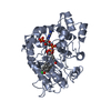
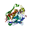
- Structure visualization
| Structure viewer | Molecule: MolmilJmol/JSmol |
|---|
- Downloads & links
-Download
| PDBx/mmCIF format | 3vxg.cif.gz | 76.6 KB | Display | PDBx/mmCIF format |
|---|---|---|---|---|
| PDB format | pdb3vxg.ent.gz | 56.3 KB | Display | PDB format |
| PDBx/mmJSON format | 3vxg.json.gz | Tree view | PDBx/mmJSON format | |
| Others |  Other downloads Other downloads |
-Validation report
| Arichive directory | https://data.pdbj.org/pub/pdb/validation_reports/vx/3vxgftp://data.pdbj.org/pub/pdb/validation_reports/vx/3vxg | HTTPS FTP |
|---|
-Related structure data
| Related structure data |  4h8nC  1vp5S C: citing same article ( S: Starting model for refinement |
|---|---|
| Similar structure data |


-Links
 PDBj
PDBj
- Assembly
| Deposited unit | 
| ||||||||
|---|---|---|---|---|---|---|---|---|---|
| 1 |
| ||||||||
| Unit cell |
|
-Components
| #1: Protein | Mass: 36865.660 Da / Num. of mol.: 1 Source method: isolated from a genetically manipulated source Source: (gene. exp.) Candida parapsilosis (yeast) / Strain: IFO 0708 / Gene: cpr-c2 / Plasmid: PET-28A / Production host:  ESCHERICHIA COLI (E. coli) / Strain (production host): ROSETTA(DE3) / References: UniProt: Q76L36, EC: 1.1.1.214 ESCHERICHIA COLI (E. coli) / Strain (production host): ROSETTA(DE3) / References: UniProt: Q76L36, EC: 1.1.1.214 |
|---|---|
| #2: Water | ChemComp-HOH / Water Mass: 18.015 Da / Num. of mol.: 213 / Source method: isolated from a natural source / Formula: H2O Mass: 18.015 Da / Num. of mol.: 213 / Source method: isolated from a natural source / Formula: H2O |
-Experimental details
-Experiment
| Experiment | Method: X-RAY DIFFRACTION / Number of used crystals: 1 |
|---|
- Sample preparation
| Crystal | Density Matthews: 1.76 Å3/Da / Density % sol: 29.98 % |
|---|---|
| Crystal grow | Temperature: 293 K / Method: vapor diffusion, sitting drop / pH: 8.1 Details: 23% PEG 3350, 0.1M TRIS-HCL, pH 8.1, VAPOR DIFFUSION, SITTING DROP, temperature 293K |
-Data collection
| Diffraction | Mean temperature: 100 K |
|---|---|
| Diffraction source | Source: SYNCHROTRON / Site: Photon Factory  / Beamline: AR-NW12A / Wavelength: 1 / Beamline: AR-NW12A / Wavelength: 1 |
| Detector | Type: ADSC QUANTUM 210 / Detector: CCD / Date: Nov 8, 2008 |
| Radiation | Protocol: SINGLE WAVELENGTH / Monochromatic (M) / Laue (L): M / Scattering type: x-ray |
| Radiation wavelength | Wavelength: 1 Å / Relative weight: 1 |
| Reflection | Resolution: 1.7→20 Å / Num. obs: 29200 / % possible obs: 99.8 % / Rmerge(I) obs: 0.044 / Net I/σ(I): 28.91 |
- Processing
| Software |
| ||||||||||||||||||||||||||||||||||||||||||||||||||||||||||||||||||||||||||||||||||||||||||||||||||||||||||||||||||||||||||||||||||||||||||||||||||||||||||||||||||||||||||
|---|---|---|---|---|---|---|---|---|---|---|---|---|---|---|---|---|---|---|---|---|---|---|---|---|---|---|---|---|---|---|---|---|---|---|---|---|---|---|---|---|---|---|---|---|---|---|---|---|---|---|---|---|---|---|---|---|---|---|---|---|---|---|---|---|---|---|---|---|---|---|---|---|---|---|---|---|---|---|---|---|---|---|---|---|---|---|---|---|---|---|---|---|---|---|---|---|---|---|---|---|---|---|---|---|---|---|---|---|---|---|---|---|---|---|---|---|---|---|---|---|---|---|---|---|---|---|---|---|---|---|---|---|---|---|---|---|---|---|---|---|---|---|---|---|---|---|---|---|---|---|---|---|---|---|---|---|---|---|---|---|---|---|---|---|---|---|---|---|---|---|---|
| Refinement | Method to determine structure: MOLECULAR REPLACEMENT Starting model: PDB ENTRY 1VP5 Resolution: 1.7→19 Å / Cor.coef. Fo:Fc: 0.956 / Cor.coef. Fo:Fc free: 0.943 / SU B: 2.354 / SU ML: 0.079 / Cross valid method: THROUGHOUT / σ(F): 0 / ESU R: 0.125 / ESU R Free: 0.118 / Stereochemistry target values: MAXIMUM LIKELIHOOD / Details: HYDROGENS HAVE BEEN ADDED IN THE RIDING POSITIONS
| ||||||||||||||||||||||||||||||||||||||||||||||||||||||||||||||||||||||||||||||||||||||||||||||||||||||||||||||||||||||||||||||||||||||||||||||||||||||||||||||||||||||||||
| Solvent computation | Ion probe radii: 0.8 Å / Shrinkage radii: 0.8 Å / VDW probe radii: 1.2 Å / Solvent model: MASK | ||||||||||||||||||||||||||||||||||||||||||||||||||||||||||||||||||||||||||||||||||||||||||||||||||||||||||||||||||||||||||||||||||||||||||||||||||||||||||||||||||||||||||
| Displacement parameters | Biso mean: 20.97 Å2
| ||||||||||||||||||||||||||||||||||||||||||||||||||||||||||||||||||||||||||||||||||||||||||||||||||||||||||||||||||||||||||||||||||||||||||||||||||||||||||||||||||||||||||
| Refinement step | Cycle: LAST / Resolution: 1.7→19 Å
| ||||||||||||||||||||||||||||||||||||||||||||||||||||||||||||||||||||||||||||||||||||||||||||||||||||||||||||||||||||||||||||||||||||||||||||||||||||||||||||||||||||||||||
| Refine LS restraints |
| ||||||||||||||||||||||||||||||||||||||||||||||||||||||||||||||||||||||||||||||||||||||||||||||||||||||||||||||||||||||||||||||||||||||||||||||||||||||||||||||||||||||||||
| LS refinement shell | Resolution: 1.7→1.74 Å / Total num. of bins used: 20
|
