 Movie
Movie Controller

[English] 日本語
 Yorodumi
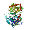
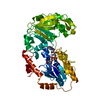
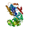
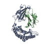
- PDB-3vue: Crystal Structure of Rice Granule bound Starch Synthase I Catalyt... -
+ Open data

- Basic information
| Entry | Database: PDB / ID: 3vue | ||||||
|---|---|---|---|---|---|---|---|
| Title | Crystal Structure of Rice Granule bound Starch Synthase I Catalytic Domain | ||||||
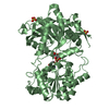
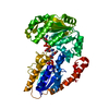
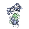
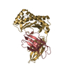
 Components Components | Granule-bound starch synthase 1, chloroplastic/amyloplastic | ||||||
 Keywords Keywords |  TRANSFERASE / Rossmann fold / glycosyltransferase TRANSFERASE / Rossmann fold / glycosyltransferase | ||||||
| Function / homology |  Function and homology informationNDP-glucose-starch glucosyltransferase / ADP-glucose-starch glucosyltransferase activity / starch biosynthetic process / amyloplast / glycogen (starch) synthase activity / IgE binding / chloroplast / ADP binding Function and homology informationNDP-glucose-starch glucosyltransferase / ADP-glucose-starch glucosyltransferase activity / starch biosynthetic process / amyloplast / glycogen (starch) synthase activity / IgE binding / chloroplast / ADP bindingSimilarity search - Function | ||||||
| Biological species |  Oryza sativa Japonica Group (Japanese rice) Oryza sativa Japonica Group (Japanese rice) | ||||||
| Method | X-RAY DIFFRACTION / SYNCHROTRON / MOLECULAR REPLACEMENT / Resolution: 2.7 Å | ||||||
 Authors Authors | Momma, M. / Fujimoto, Z. | ||||||
 Citation Citation | Journal: Biosci.Biotechnol.Biochem. / Year: 2012 Title: Interdomain Disulfide Bridge in the Rice Granule Bound Starch Synthase I Catalytic Domain as Elucidated by X-Ray Structure Analysis Authors: Momma, M. / Fujimoto, Z. | ||||||
| History |
|
- Structure visualization
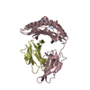
| Structure viewer | Molecule: MolmilJmol/JSmol |
|---|
- Downloads & links
-Download
| PDBx/mmCIF format | 3vue.cif.gz | 110.6 KB | Display | PDBx/mmCIF format |
|---|---|---|---|---|
| PDB format | pdb3vue.ent.gz | 84.2 KB | Display | PDB format |
| PDBx/mmJSON format | 3vue.json.gz | Tree view | PDBx/mmJSON format | |
| Others |  Other downloads Other downloads |
-Validation report
| Arichive directory | https://data.pdbj.org/pub/pdb/validation_reports/vu/3vueftp://data.pdbj.org/pub/pdb/validation_reports/vu/3vue | HTTPS FTP |
|---|
-Related structure data
| Related structure data |  3vufC 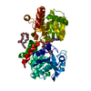 3guhS C: citing same article ( S: Starting model for refinement |
|---|---|
| Similar structure data |

-Links
 PDBj
PDBj
- Assembly
| Deposited unit | 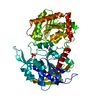
| ||||||||
|---|---|---|---|---|---|---|---|---|---|
| 1 |
| ||||||||
| Unit cell |
|
-Components
| #1: Protein | Mass: 59354.000 Da / Num. of mol.: 1 / Fragment: catalytic domain, UNP residues 83-609 Source method: isolated from a genetically manipulated source Source: (gene. exp.) Oryza sativa Japonica Group (Japanese rice)Strain: japonica cultivar-group Gene: WAXY, WX, WX-B, Os06g0133000, LOC_Os06g04200, 134P10.7, P0679C08.19 Plasmid: pET45b / Production host:  Escherichia coli (E. coli) / Strain (production host): BL-21(DE3) gold Escherichia coli (E. coli) / Strain (production host): BL-21(DE3) goldReferences: UniProt: Q0DEV5, NDP-glucose-starch glucosyltransferase | ||
|---|---|---|---|
| #2: Chemical | ChemComp-SO4 / Sulfate  Mass: 96.063 Da / Num. of mol.: 4 / Source method: obtained synthetically / Formula: SO4 Mass: 96.063 Da / Num. of mol.: 4 / Source method: obtained synthetically / Formula: SO4#3: Water | ChemComp-HOH / | Water Mass: 18.015 Da / Num. of mol.: 61 / Source method: isolated from a natural source / Formula: H2O Mass: 18.015 Da / Num. of mol.: 61 / Source method: isolated from a natural source / Formula: H2O |
-Experimental details
-Experiment
| Experiment | Method: X-RAY DIFFRACTION / Number of used crystals: 1 |
|---|
- Sample preparation
| Crystal | Density Matthews: 2.51 Å3/Da / Density % sol: 50.99 % |
|---|---|
| Crystal grow | Temperature: 293 K / Method: vapor diffusion, sitting drop / pH: 7.5 Details: 1.5M Lithium Sulfate, 0.1M HEPES, pH 7.5, VAPOR DIFFUSION, SITTING DROP, temperature 293K |
-Data collection
| Diffraction | Mean temperature: 95 K |
|---|---|
| Diffraction source | Source: SYNCHROTRON / Site: Photon Factory  / Beamline: AR-NW12A / Wavelength: 1 Å / Beamline: AR-NW12A / Wavelength: 1 Å |
| Detector | Type: ADSC QUANTUM 210 / Detector: CCD / Date: Jan 1, 2009 |
| Radiation | Monochromator: Si 111 / Protocol: SINGLE WAVELENGTH / Monochromatic (M) / Laue (L): M / Scattering type: x-ray |
| Radiation wavelength | Wavelength: 1 Å / Relative weight: 1 |
| Reflection | Resolution: 2.7→50 Å / Num. obs: 17202 / % possible obs: 99.3 % / Redundancy: 13.5 % / Biso Wilson estimate: 78.6 Å2 / Rmerge(I) obs: 0.046 / Net I/σ(I): 27.5 |
| Reflection shell | Resolution: 2.7→2.8 Å / Redundancy: 13.9 % / Rmerge(I) obs: 0.37 / Mean I/σ(I) obs: 7 / Num. unique all: 1681 / % possible all: 99.9 |
- Processing
| Software |
| |||||||||||||||||||||||||||||||||||||||||||||||||||||||||||||||||
|---|---|---|---|---|---|---|---|---|---|---|---|---|---|---|---|---|---|---|---|---|---|---|---|---|---|---|---|---|---|---|---|---|---|---|---|---|---|---|---|---|---|---|---|---|---|---|---|---|---|---|---|---|---|---|---|---|---|---|---|---|---|---|---|---|---|---|
| Refinement | Method to determine structure: MOLECULAR REPLACEMENT Starting model: 3GUH Resolution: 2.7→50 Å / Cor.coef. Fo:Fc: 0.937 / Cor.coef. Fo:Fc free: 0.897 / SU B: 11.367 / SU ML: 0.235 / Cross valid method: THROUGHOUT / ESU R: 1.792 / ESU R Free: 0.365 / Stereochemistry target values: MAXIMUM LIKELIHOOD / Details: HYDROGENS HAVE BEEN ADDED IN THE RIDING POSITIONS
| |||||||||||||||||||||||||||||||||||||||||||||||||||||||||||||||||
| Solvent computation | Ion probe radii: 0.8 Å / Shrinkage radii: 0.8 Å / VDW probe radii: 1.4 Å / Solvent model: MASK | |||||||||||||||||||||||||||||||||||||||||||||||||||||||||||||||||
| Displacement parameters | Biso mean: 61.896 Å2 | |||||||||||||||||||||||||||||||||||||||||||||||||||||||||||||||||
| Refinement step | Cycle: LAST / Resolution: 2.7→50 Å
| |||||||||||||||||||||||||||||||||||||||||||||||||||||||||||||||||
| Refine LS restraints |
| |||||||||||||||||||||||||||||||||||||||||||||||||||||||||||||||||
| LS refinement shell | Resolution: 2.7→2.77 Å / Total num. of bins used: 20
|
