 Movie
Movie Controller

[English] 日本語
 Yorodumi
- PDB-3vu2: Structure of the Starch Branching Enzyme I (BEI) complexed with m... -
+ Open data

- Basic information
| Entry | Database: PDB / ID: 3vu2 | |||||||||
|---|---|---|---|---|---|---|---|---|---|---|
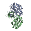
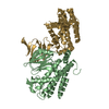
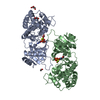
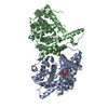
| Title | Structure of the Starch Branching Enzyme I (BEI) complexed with maltopentaose from Oryza sativa L | |||||||||
 Components Components | 1,4-alpha-glucan-branching enzyme, chloroplastic/amyloplastic | |||||||||
 Keywords Keywords |  TRANSFERASE / Carbohydrate-binding module 48 TRANSFERASE / Carbohydrate-binding module 48 | |||||||||
| Function / homology |  Function and homology information Function and homology informationstarch metabolic process / starch biosynthetic process / amyloplast / 1,4-alpha-glucan branching enzyme / 1,4-alpha-glucan branching enzyme activity (using a glucosylated glycogenin as primer for glycogen synthesis) / 1,4-alpha-glucan branching enzyme activity / cation binding / starch catabolic process / glycogen biosynthetic process / hydrolase activity, hydrolyzing O-glycosyl compounds ...starch metabolic process / starch biosynthetic process / amyloplast / 1,4-alpha-glucan branching enzyme / 1,4-alpha-glucan branching enzyme activity (using a glucosylated glycogenin as primer for glycogen synthesis) / 1,4-alpha-glucan branching enzyme activity / cation binding / starch catabolic process / glycogen biosynthetic process / hydrolase activity, hydrolyzing O-glycosyl compounds / chloroplast / carbohydrate metabolic process / cytoplasmSimilarity search - Function | |||||||||
| Biological species |  Oryza sativa Japonica Group (Japanese rice) Oryza sativa Japonica Group (Japanese rice) | |||||||||
| Method | X-RAY DIFFRACTION / SYNCHROTRON / MOLECULAR REPLACEMENT / Resolution: 2.23 Å | |||||||||
 Authors Authors | Chaen, K. / Kakuta, Y. / Kimura, M. | |||||||||
 Citation Citation | Journal: Biochem.Biophys.Res.Commun. / Year: 2012 Title: Crystal structure of the rice branching enzyme I (BEI) in complex with maltopentaose. Authors: Chaen, K. / Noguchi, J. / Omori, T. / Kakuta, Y. / Kimura, M. | |||||||||
| History |
|
- Structure visualization
| Structure viewer | Molecule: MolmilJmol/JSmol |
|---|
- Downloads & links
-Download
| PDBx/mmCIF format | 3vu2.cif.gz | 305.4 KB | Display | PDBx/mmCIF format |
|---|---|---|---|---|
| PDB format | pdb3vu2.ent.gz | 246.9 KB | Display | PDB format |
| PDBx/mmJSON format | 3vu2.json.gz | Tree view | PDBx/mmJSON format | |
| Others |  Other downloads Other downloads |
-Validation report
| Arichive directory | https://data.pdbj.org/pub/pdb/validation_reports/vu/3vu2ftp://data.pdbj.org/pub/pdb/validation_reports/vu/3vu2 | HTTPS FTP |
|---|
-Related structure data
| Related structure data | |
|---|---|
| Similar structure data |


-Links
 PDBj
PDBj


- Assembly
| Deposited unit | 
| ||||||||
|---|---|---|---|---|---|---|---|---|---|
| 1 | 
| ||||||||
| 2 | 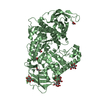
| ||||||||
| Unit cell |
|
-Components
| #1: Protein | Mass: 81201.578 Da / Num. of mol.: 2 / Fragment: UNP RESIDUES 66-767 / Mutation: E399Q Source method: isolated from a genetically manipulated source Source: (gene. exp.) Oryza sativa Japonica Group (Japanese rice)Gene: SBE1, RBE1, Os06g0726400, LOC_Os06g51084, P0017G10.8-1, P0017G10.8-2, P0548E04.28-1, P0548E04.28-2 Production host:  Escherichia coli (E. coli) Escherichia coli (E. coli)References: UniProt: Q01401, 1,4-alpha-glucan branching enzyme#2: Polysaccharide | 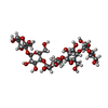  , Oligosaccharide / Class: Substrate analog / Mass: 828.719 Da / Num. of mol.: 2 , Oligosaccharide / Class: Substrate analog / Mass: 828.719 Da / Num. of mol.: 2Source method: isolated from a genetically manipulated source Details: oligosaccharide / References: alpha-maltopentaose #3: Sugar | ChemComp-BGC / Glucose  Type: D-saccharide, beta linking / Mass: 180.156 Da / Num. of mol.: 4 Type: D-saccharide, beta linking / Mass: 180.156 Da / Num. of mol.: 4Source method: isolated from a genetically manipulated source Formula: C6H12O6 #4: Chemical | ChemComp-GOL / Glycerol  Mass: 92.094 Da / Num. of mol.: 5 / Source method: obtained synthetically / Formula: C3H8O3 Mass: 92.094 Da / Num. of mol.: 5 / Source method: obtained synthetically / Formula: C3H8O3#5: Water | ChemComp-HOH / | Water Mass: 18.015 Da / Num. of mol.: 787 / Source method: isolated from a natural source / Formula: H2O Mass: 18.015 Da / Num. of mol.: 787 / Source method: isolated from a natural source / Formula: H2O |
|---|
-Experimental details
-Experiment
| Experiment | Method: X-RAY DIFFRACTION |
|---|
- Sample preparation
| Crystal | Density Matthews: 2.57 Å3/Da / Density % sol: 52.06 % |
|---|
-Data collection
| Diffraction | Mean temperature: 100 K |
|---|---|
| Diffraction source | Source: SYNCHROTRON / Site: SPring-8  / Beamline: BL38B1 / Wavelength: 1 Å / Beamline: BL38B1 / Wavelength: 1 Å |
| Detector | Type: ADSC QUANTUM 315 / Detector: CCD |
| Radiation | Monochromator: Si 111 CHANNEL / Protocol: SINGLE WAVELENGTH / Monochromatic (M) / Laue (L): M / Scattering type: x-ray |
| Radiation wavelength | Wavelength: 1 Å / Relative weight: 1 |
| Reflection | Resolution: 2.2→50 Å / Num. all: 72156 / Num. obs: 72156 / % possible obs: 88.5 % / Observed criterion σ(F): 0 / Observed criterion σ(I): 0 |
- Processing
| Software |
| ||||||||||||||||||||||||||||||||||||||||||||||||||||||||||||||||||||||||||||||||||||||||||||||||||||||||||||||||||||||||||||||||||||||||||||||||||||||||||||||||||||||||||
|---|---|---|---|---|---|---|---|---|---|---|---|---|---|---|---|---|---|---|---|---|---|---|---|---|---|---|---|---|---|---|---|---|---|---|---|---|---|---|---|---|---|---|---|---|---|---|---|---|---|---|---|---|---|---|---|---|---|---|---|---|---|---|---|---|---|---|---|---|---|---|---|---|---|---|---|---|---|---|---|---|---|---|---|---|---|---|---|---|---|---|---|---|---|---|---|---|---|---|---|---|---|---|---|---|---|---|---|---|---|---|---|---|---|---|---|---|---|---|---|---|---|---|---|---|---|---|---|---|---|---|---|---|---|---|---|---|---|---|---|---|---|---|---|---|---|---|---|---|---|---|---|---|---|---|---|---|---|---|---|---|---|---|---|---|---|---|---|---|---|---|---|
| Refinement | Method to determine structure: MOLECULAR REPLACEMENT / Resolution: 2.23→37.22 Å / Cor.coef. Fo:Fc: 0.934 / Cor.coef. Fo:Fc free: 0.897 / SU B: 8.337 / SU ML: 0.206 / Cross valid method: THROUGHOUT / ESU R: 0.447 / ESU R Free: 0.291 / Stereochemistry target values: MAXIMUM LIKELIHOOD / Details: HYDROGENS HAVE BEEN ADDED IN THE RIDING POSITIONS
| ||||||||||||||||||||||||||||||||||||||||||||||||||||||||||||||||||||||||||||||||||||||||||||||||||||||||||||||||||||||||||||||||||||||||||||||||||||||||||||||||||||||||||
| Solvent computation | Ion probe radii: 0.8 Å / Shrinkage radii: 0.8 Å / VDW probe radii: 1.4 Å / Solvent model: MASK | ||||||||||||||||||||||||||||||||||||||||||||||||||||||||||||||||||||||||||||||||||||||||||||||||||||||||||||||||||||||||||||||||||||||||||||||||||||||||||||||||||||||||||
| Displacement parameters | Biso mean: 44.47 Å2
| ||||||||||||||||||||||||||||||||||||||||||||||||||||||||||||||||||||||||||||||||||||||||||||||||||||||||||||||||||||||||||||||||||||||||||||||||||||||||||||||||||||||||||
| Refinement step | Cycle: LAST / Resolution: 2.23→37.22 Å
| ||||||||||||||||||||||||||||||||||||||||||||||||||||||||||||||||||||||||||||||||||||||||||||||||||||||||||||||||||||||||||||||||||||||||||||||||||||||||||||||||||||||||||
| Refine LS restraints |
| ||||||||||||||||||||||||||||||||||||||||||||||||||||||||||||||||||||||||||||||||||||||||||||||||||||||||||||||||||||||||||||||||||||||||||||||||||||||||||||||||||||||||||
| LS refinement shell | Resolution: 2.234→2.292 Å / Total num. of bins used: 20
|
