 Movie
Movie Controller

[English] 日本語
 Yorodumi
- PDB-3vtw: Crystal structure of T7-tagged Optineurin LIR-fused human LC3B_2-119 -
+ Open data

- Basic information
| Entry | Database: PDB / ID: 3vtw | ||||||
|---|---|---|---|---|---|---|---|
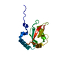
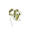
| Title | Crystal structure of T7-tagged Optineurin LIR-fused human LC3B_2-119 | ||||||
 Components Components | Optineurin, microtubule-associated proteins 1A/1B light chain 3B | ||||||
 Keywords Keywords | PROTEIN BINDING / ubiquitin-like fold / Autophagy | ||||||
| Function / homology |  Function and homology information Function and homology informationSARS-CoV-2 modulates autophagy / parkin-mediated stimulation of mitophagy in response to mitochondrial depolarization / Golgi ribbon formation / ceramide binding / negative regulation of receptor recycling / cell death / protein localization to Golgi apparatus / positive regulation of xenophagy / Golgi to plasma membrane protein transport / phosphatidylethanolamine binding ...SARS-CoV-2 modulates autophagy / parkin-mediated stimulation of mitophagy in response to mitochondrial depolarization / Golgi ribbon formation / ceramide binding / negative regulation of receptor recycling / cell death / protein localization to Golgi apparatus / positive regulation of xenophagy / Golgi to plasma membrane protein transport / phosphatidylethanolamine binding / cellular response to nitrogen starvation / Translation of Replicase and Assembly of the Replication Transcription Complex / TBC/RABGAPs / regulation of canonical NF-kappaB signal transduction / TNFR1-induced proapoptotic signaling / K63-linked polyubiquitin modification-dependent protein binding / Macroautophagy / Receptor Mediated Mitophagy / Golgi organization / axoneme / autophagosome maturation / autophagosome membrane / organelle membrane / mitophagy / autophagosome assembly / polyubiquitin modification-dependent protein binding / autophagosome / endomembrane system / cellular response to unfolded protein / positive regulation of autophagy / negative regulation of canonical NF-kappaB signal transduction / PINK1-PRKN Mediated Mitophagy / Pexophagy / cellular response to starvation / TNFR1-induced NF-kappa-B signaling pathway / mitochondrial membrane / Regulation of TNFR1 signaling / macroautophagy / trans-Golgi network / autophagy / recycling endosome membrane / KEAP1-NFE2L2 pathway / Regulation of PLK1 Activity at G2/M Transition / protein-macromolecule adaptor activity / Translation of Replicase and Assembly of the Replication Transcription Complex / cytoplasmic vesicle / microtubule binding / microtubule / defense response to Gram-negative bacterium / Golgi membrane / intracellular membrane-bounded organelle / innate immune response / ubiquitin protein ligase binding / perinuclear region of cytoplasm / Golgi apparatus / signal transduction / mitochondrion / nucleoplasm / identical protein binding / metal ion binding / nucleus / cytosol / cytoplasmSimilarity search - Function | ||||||
| Biological species |  Homo sapiens (human) Homo sapiens (human) | ||||||
| Method | X-RAY DIFFRACTION / SYNCHROTRON / MOLECULAR REPLACEMENT / Resolution: 2.52 Å | ||||||
 Authors Authors | Suzuki, H. / Kawasaki, M. / Kato, R. / Wakatsuki, S. | ||||||
 Citation Citation | Journal: Biochem.J. / Year: 2013 Title: Structural basis for phosphorylation-triggered autophagic clearance of Salmonella Authors: Rogov, V.V. / Suzuki, H. / Fiskin, E. / Wild, P. / Kniss, A. / Rozenknop, A. / Kato, R. / Kawasaki, M. / McEwan, D.G. / Lohr, F. / Guntert, P. / Dikic, I. / Wakatsuki, S. / Dotsch, V. | ||||||
| History |
|
- Structure visualization
| Structure viewer | Molecule: MolmilJmol/JSmol |
|---|
- Downloads & links
-Download
| PDBx/mmCIF format | 3vtw.cif.gz | 168.1 KB | Display | PDBx/mmCIF format |
|---|---|---|---|---|
| PDB format | pdb3vtw.ent.gz | 134.4 KB | Display | PDB format |
| PDBx/mmJSON format | 3vtw.json.gz | Tree view | PDBx/mmJSON format | |
| Others |  Other downloads Other downloads |
-Validation report
| Arichive directory | https://data.pdbj.org/pub/pdb/validation_reports/vt/3vtwftp://data.pdbj.org/pub/pdb/validation_reports/vt/3vtw | HTTPS FTP |
|---|
-Related structure data
| Related structure data |  2lueC 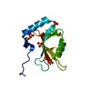 3vtuSC 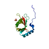 3vtvC S: Starting model for refinement C: citing same article ( |
|---|---|
| Similar structure data |


-Links
 PDBj
PDBj


- Assembly
| Deposited unit | 
| ||||||||
|---|---|---|---|---|---|---|---|---|---|
| 1 | 
| ||||||||
| 2 | 
| ||||||||
| 3 | 
| ||||||||
| Unit cell |
|
-Components
| #1: Protein | / Autophagy-related protein LC3 B / Autophagy-related ubiquitin-like modifier LC3 B / MAP1 light ...Autophagy-related protein LC3 B / Autophagy-related ubiquitin-like modifier LC3 B / MAP1 light chain 3-like protein 2 / MAP1A/MAP1B light chain 3 B / MAP1A/MAP1B LC3 B / Microtubule-associated protein 1 light chain 3 beta Mass: 17175.408 Da / Num. of mol.: 3 / Fragment: UNP RESIDUES 170-181, RESIDUES 2-119 / Mutation: S170E, S171E, S173E, S174E, S177E Source method: isolated from a genetically manipulated source Details: The fusion protein of Optineurin LIR (RESIDUES 170-181) and Microtubule-associated proteins 1A/1B light chain 3B (RESIDUES 2-119) Source: (gene. exp.) Homo sapiens (human) / Gene: MAP1LC3B, MAP1ALC3 / Plasmid: pET28a / Production host:  Escherichia coli (E. coli) / Strain (production host): BL21(DE3) / References: UniProt: Q96CV9, UniProt: Q9GZQ8 Escherichia coli (E. coli) / Strain (production host): BL21(DE3) / References: UniProt: Q96CV9, UniProt: Q9GZQ8#2: Chemical | ChemComp-SO4 / Sulfate  Mass: 96.063 Da / Num. of mol.: 4 / Source method: obtained synthetically / Formula: SO4 Mass: 96.063 Da / Num. of mol.: 4 / Source method: obtained synthetically / Formula: SO4#3: Water | ChemComp-HOH / | Water Mass: 18.015 Da / Num. of mol.: 85 / Source method: isolated from a natural source / Formula: H2O Mass: 18.015 Da / Num. of mol.: 85 / Source method: isolated from a natural source / Formula: H2O |
|---|
-Experimental details
-Experiment
| Experiment | Method: X-RAY DIFFRACTION / Number of used crystals: 1 |
|---|
- Sample preparation
| Crystal | Density Matthews: 2.85 Å3/Da / Density % sol: 56.84 % |
|---|---|
| Crystal grow | Temperature: 289 K / Method: vapor diffusion, hanging drop / pH: 5.6 Details: 2.0M Ammonium sulfate, 0.05M Tri-sodium citrate, 0.1M Potassium sodium tartrate, 5% Glycerol, pH 5.6, VAPOR DIFFUSION, HANGING DROP, temperature 289K |
-Data collection
| Diffraction | Mean temperature: 100 K |
|---|---|
| Diffraction source | Source: SYNCHROTRON / Site: Photon Factory  / Beamline: BL-5A / Wavelength: 1 Å / Beamline: BL-5A / Wavelength: 1 Å |
| Detector | Type: ADSC QUANTUM 210r / Detector: CCD / Date: Jul 5, 2011 |
| Radiation | Monochromator: Numerical link type Si(111) double crystal monochromator Protocol: SINGLE WAVELENGTH / Monochromatic (M) / Laue (L): M / Scattering type: x-ray |
| Radiation wavelength | Wavelength: 1 Å / Relative weight: 1 |
| Reflection | Resolution: 2.52→49.92 Å / Num. obs: 20055 / % possible obs: 97.5 % / Rmerge(I) obs: 0.128 / Net I/σ(I): 12.9 |
| Reflection shell | Resolution: 2.52→2.66 Å / Rmerge(I) obs: 0.701 / Mean I/σ(I) obs: 3 / Num. unique all: 2828 / % possible all: 95.9 |
- Processing
| Software |
| ||||||||||||||||||||||||||||||||||||||||||||||||||||||||||||||||||||||||||||||||||||||||||||||||||||||||||||||||||||||||||||||||||||||||||||||||||||||||||||||||||||||||||
|---|---|---|---|---|---|---|---|---|---|---|---|---|---|---|---|---|---|---|---|---|---|---|---|---|---|---|---|---|---|---|---|---|---|---|---|---|---|---|---|---|---|---|---|---|---|---|---|---|---|---|---|---|---|---|---|---|---|---|---|---|---|---|---|---|---|---|---|---|---|---|---|---|---|---|---|---|---|---|---|---|---|---|---|---|---|---|---|---|---|---|---|---|---|---|---|---|---|---|---|---|---|---|---|---|---|---|---|---|---|---|---|---|---|---|---|---|---|---|---|---|---|---|---|---|---|---|---|---|---|---|---|---|---|---|---|---|---|---|---|---|---|---|---|---|---|---|---|---|---|---|---|---|---|---|---|---|---|---|---|---|---|---|---|---|---|---|---|---|---|---|---|
| Refinement | Method to determine structure: MOLECULAR REPLACEMENT Starting model: PDB ENTRY 3VTU Resolution: 2.52→49.92 Å / Cor.coef. Fo:Fc: 0.927 / Cor.coef. Fo:Fc free: 0.877 / SU B: 21.796 / SU ML: 0.217 / Cross valid method: THROUGHOUT / ESU R: 0.395 / ESU R Free: 0.298 / Stereochemistry target values: MAXIMUM LIKELIHOOD / Details: HYDROGENS HAVE BEEN ADDED IN THE RIDING POSITIONS
| ||||||||||||||||||||||||||||||||||||||||||||||||||||||||||||||||||||||||||||||||||||||||||||||||||||||||||||||||||||||||||||||||||||||||||||||||||||||||||||||||||||||||||
| Solvent computation | Ion probe radii: 0.8 Å / Shrinkage radii: 0.8 Å / VDW probe radii: 1.4 Å / Solvent model: MASK | ||||||||||||||||||||||||||||||||||||||||||||||||||||||||||||||||||||||||||||||||||||||||||||||||||||||||||||||||||||||||||||||||||||||||||||||||||||||||||||||||||||||||||
| Displacement parameters | Biso mean: 36.628 Å2
| ||||||||||||||||||||||||||||||||||||||||||||||||||||||||||||||||||||||||||||||||||||||||||||||||||||||||||||||||||||||||||||||||||||||||||||||||||||||||||||||||||||||||||
| Refinement step | Cycle: LAST / Resolution: 2.52→49.92 Å
| ||||||||||||||||||||||||||||||||||||||||||||||||||||||||||||||||||||||||||||||||||||||||||||||||||||||||||||||||||||||||||||||||||||||||||||||||||||||||||||||||||||||||||
| Refine LS restraints |
| ||||||||||||||||||||||||||||||||||||||||||||||||||||||||||||||||||||||||||||||||||||||||||||||||||||||||||||||||||||||||||||||||||||||||||||||||||||||||||||||||||||||||||
| LS refinement shell | Resolution: 2.52→2.585 Å / Total num. of bins used: 20
| ||||||||||||||||||||||||||||||||||||||||||||||||||||||||||||||||||||||||||||||||||||||||||||||||||||||||||||||||||||||||||||||||||||||||||||||||||||||||||||||||||||||||||
| Refinement TLS params. | Method: refined / Refine-ID: X-RAY DIFFRACTION
| ||||||||||||||||||||||||||||||||||||||||||||||||||||||||||||||||||||||||||||||||||||||||||||||||||||||||||||||||||||||||||||||||||||||||||||||||||||||||||||||||||||||||||
| Refinement TLS group |
|
