 Movie
Movie Controller


+ Open data

- Basic information
| Entry | Database: PDB / ID: 3vrh | ||||||
|---|---|---|---|---|---|---|---|
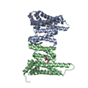
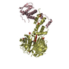
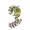
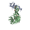
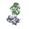
| Title | Crystal structure of ph0300 | ||||||
 Components Components | Putative uncharacterized protein PH0300 | ||||||
 Keywords Keywords |  RNA BINDING PROTEIN / ATPase / tRNA modification enzyme / thiolation RNA BINDING PROTEIN / ATPase / tRNA modification enzyme / thiolation | ||||||
| Function / homology |  Function and homology informationTransferases; Transferring sulfur-containing groups; Sulfurtransferases / tRNA thio-modification / tRNA wobble uridine modification / 4 iron, 4 sulfur cluster binding / transferase activity / tRNA binding / ATP binding / metal ion binding Function and homology informationTransferases; Transferring sulfur-containing groups; Sulfurtransferases / tRNA thio-modification / tRNA wobble uridine modification / 4 iron, 4 sulfur cluster binding / transferase activity / tRNA binding / ATP binding / metal ion bindingSimilarity search - Function | ||||||
| Biological species |   Pyrococcus horikoshii (archaea) Pyrococcus horikoshii (archaea) | ||||||
| Method | X-RAY DIFFRACTION / SYNCHROTRON / SAD / Resolution: 2.1 Å | ||||||
 Authors Authors | Nakagawa, H. / Kuratani, M. / Goto-Ito, S. / Ito, T. / Sekine, S.I. / Yokoyama, S. | ||||||
 Citation Citation | Journal: Proteins / Year: 2013 Title: Crystallographic and mutational studies on the tRNA thiouridine synthetase TtuA. Authors: Nakagawa, H. / Kuratani, M. / Goto-Ito, S. / Ito, T. / Katsura, K. / Terada, T. / Shirouzu, M. / Sekine, S.I. / Shigi, N. / Yokoyama, S. | ||||||
| History |
|
- Structure visualization
| Structure viewer | Molecule: MolmilJmol/JSmol |
|---|
- Downloads & links
-Download
| PDBx/mmCIF format | 3vrh.cif.gz | 70.4 KB | Display | PDBx/mmCIF format |
|---|---|---|---|---|
| PDB format | pdb3vrh.ent.gz | 55.9 KB | Display | PDB format |
| PDBx/mmJSON format | 3vrh.json.gz | Tree view | PDBx/mmJSON format | |
| Others |  Other downloads Other downloads |
-Validation report
| Arichive directory | https://data.pdbj.org/pub/pdb/validation_reports/vr/3vrhftp://data.pdbj.org/pub/pdb/validation_reports/vr/3vrh | HTTPS FTP |
|---|
-Related structure data
| Similar structure data |
|---|


-Links
 PDBj
PDBj- Assembly
| Deposited unit | 
| ||||||||
|---|---|---|---|---|---|---|---|---|---|
| 1 | 
| ||||||||
| Unit cell |
|
-Components
| #1: Protein | Mass: 35691.672 Da / Num. of mol.: 1 Source method: isolated from a genetically manipulated source Source: (gene. exp.) Pyrococcus horikoshii (archaea) / Strain: OT-3 / Gene: PH0300 / Production host:  Escherichia coli (E. coli) / References: UniProt: O58038 Escherichia coli (E. coli) / References: UniProt: O58038 | ||||
|---|---|---|---|---|---|
| #2: Chemical |   Mass: 65.409 Da / Num. of mol.: 2 / Source method: obtained synthetically / Formula: Zn Mass: 65.409 Da / Num. of mol.: 2 / Source method: obtained synthetically / Formula: Zn#3: Chemical | ChemComp-BCN / | Bicine  Mass: 163.172 Da / Num. of mol.: 1 / Source method: obtained synthetically / Formula: C6H13NO4 / Comment: pH buffer*YM Mass: 163.172 Da / Num. of mol.: 1 / Source method: obtained synthetically / Formula: C6H13NO4 / Comment: pH buffer*YM#4: Water | ChemComp-HOH / | Water Mass: 18.015 Da / Num. of mol.: 80 / Source method: isolated from a natural source / Formula: H2O Mass: 18.015 Da / Num. of mol.: 80 / Source method: isolated from a natural source / Formula: H2O |
-Experimental details
-Experiment
| Experiment | Method: X-RAY DIFFRACTION / Number of used crystals: 1 |
|---|
- Sample preparation
| Crystal | Density Matthews: 2.21 Å3/Da / Density % sol: 44.39 % Description: The entry contains Friedel pairs in F_Plus/Minus columns |
|---|---|
| Crystal grow | Temperature: 293 K / Method: vapor diffusion, sitting drop / pH: 9 Details: 12% PEG 3350, 0.2 M NaCl, 10% N,N-dimethylformamide, 0.1 M Bicine-NaOH, pH 9, VAPOR DIFFUSION, SITTING DROP, temperature 293K |
-Data collection
| Diffraction | Mean temperature: 70 K | |||||||||||||||||||||||||||||||||
|---|---|---|---|---|---|---|---|---|---|---|---|---|---|---|---|---|---|---|---|---|---|---|---|---|---|---|---|---|---|---|---|---|---|---|
| Diffraction source | Source: SYNCHROTRON / Site: SPring-8  / Beamline: BL32XU / Wavelength: 1 Å / Beamline: BL32XU / Wavelength: 1 Å | |||||||||||||||||||||||||||||||||
| Detector | Type: RAYONIX MX225HE / Detector: CCD / Date: Jan 1, 2010 | |||||||||||||||||||||||||||||||||
| Radiation | Monochromator: silicon / Protocol: SINGLE WAVELENGTH / Monochromatic (M) / Laue (L): M / Scattering type: x-ray | |||||||||||||||||||||||||||||||||
| Radiation wavelength | Wavelength: 1 Å / Relative weight: 1 | |||||||||||||||||||||||||||||||||
| Reflection | Resolution: 2.1→50 Å / Num. all: 19954 / Num. obs: 19923 / % possible obs: 99.9 % / Observed criterion σ(F): 5 / Observed criterion σ(I): 25.1 | |||||||||||||||||||||||||||||||||
| Reflection shell |
|
- Processing
| Software |
| ||||||||||||||||||||||||||||||||||||||||||||||||||||||||||||||||||||||||||||||||||||
|---|---|---|---|---|---|---|---|---|---|---|---|---|---|---|---|---|---|---|---|---|---|---|---|---|---|---|---|---|---|---|---|---|---|---|---|---|---|---|---|---|---|---|---|---|---|---|---|---|---|---|---|---|---|---|---|---|---|---|---|---|---|---|---|---|---|---|---|---|---|---|---|---|---|---|---|---|---|---|---|---|---|---|---|---|---|
| Refinement | Method to determine structure: SAD / Resolution: 2.1→46.236 Å / SU ML: 0.31 / σ(F): 1.91 / Phase error: 24.03 / Stereochemistry target values: ML Details: The entry contains Friedel pairs in F_Plus/Minus columns
| ||||||||||||||||||||||||||||||||||||||||||||||||||||||||||||||||||||||||||||||||||||
| Solvent computation | Shrinkage radii: 0.98 Å / VDW probe radii: 1.2 Å / Solvent model: FLAT BULK SOLVENT MODEL / Bsol: 38.677 Å2 / ksol: 0.333 e/Å3 | ||||||||||||||||||||||||||||||||||||||||||||||||||||||||||||||||||||||||||||||||||||
| Displacement parameters |
| ||||||||||||||||||||||||||||||||||||||||||||||||||||||||||||||||||||||||||||||||||||
| Refinement step | Cycle: LAST / Resolution: 2.1→46.236 Å
| ||||||||||||||||||||||||||||||||||||||||||||||||||||||||||||||||||||||||||||||||||||
| Refine LS restraints |
| ||||||||||||||||||||||||||||||||||||||||||||||||||||||||||||||||||||||||||||||||||||
| LS refinement shell | Refine-ID: X-RAY DIFFRACTION / Total num. of bins used: 13
|
