 Movie
Movie Controller

[English] 日本語
 Yorodumi
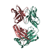
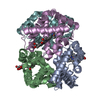
- PDB-3vrf: The crystal structure of hemoglobin from woolly mammoth in the ca... -
+ Open data

- Basic information
| Entry | Database: PDB / ID: 3vrf | ||||||
|---|---|---|---|---|---|---|---|
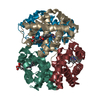
| Title | The crystal structure of hemoglobin from woolly mammoth in the carbonmonoxy forms | ||||||
 Components Components |
| ||||||
 Keywords Keywords |  OXYGEN TRANSPORT / woolly mammoth hemoglobin / OXYGEN STORAGE/TRANSPORT OXYGEN TRANSPORT / woolly mammoth hemoglobin / OXYGEN STORAGE/TRANSPORT | ||||||
| Function / homology |  Function and homology informationhaptoglobin binding / hemoglobin alpha binding / haptoglobin-hemoglobin complex / organic acid binding / hemoglobin complex / hydrogen peroxide catabolic process / oxygen carrier activity / peroxidase activity / oxygen binding / blood microparticle ...haptoglobin binding / hemoglobin alpha binding / haptoglobin-hemoglobin complex / organic acid binding / hemoglobin complex / hydrogen peroxide catabolic process / oxygen carrier activity / peroxidase activity / oxygen binding / blood microparticle / iron ion binding / heme binding / metal ion binding Function and homology informationhaptoglobin binding / hemoglobin alpha binding / haptoglobin-hemoglobin complex / organic acid binding / hemoglobin complex / hydrogen peroxide catabolic process / oxygen carrier activity / peroxidase activity / oxygen binding / blood microparticle ...haptoglobin binding / hemoglobin alpha binding / haptoglobin-hemoglobin complex / organic acid binding / hemoglobin complex / hydrogen peroxide catabolic process / oxygen carrier activity / peroxidase activity / oxygen binding / blood microparticle / iron ion binding / heme binding / metal ion bindingSimilarity search - Function | ||||||
| Biological species |  Mammuthus primigenius (woolly mammoth) Mammuthus primigenius (woolly mammoth) | ||||||
| Method | X-RAY DIFFRACTION / SYNCHROTRON / MOLECULAR REPLACEMENT / Resolution: 1.55 Å | ||||||
 Authors Authors | Noguchi, H. / Campbell, K.L. / Ho, C. / Park, S.-Y. / Tame, J.R.H. | ||||||
 Citation Citation | Journal: Acta Crystallogr.,Sect.D / Year: 2012 Title: Structures of haemoglobin from woolly mammoth in liganded and unliganded states. Authors: Noguchi, H. / Campbell, K.L. / Ho, C. / Unzai, S. / Park, S.-Y. / Tame, J.R.H. | ||||||
| History |
|
- Structure visualization
| Structure viewer | Molecule: MolmilJmol/JSmol |
|---|
- Downloads & links
-Download
| PDBx/mmCIF format | 3vrf.cif.gz | 78.2 KB | Display | PDBx/mmCIF format |
|---|---|---|---|---|
| PDB format | pdb3vrf.ent.gz | 57.6 KB | Display | PDB format |
| PDBx/mmJSON format | 3vrf.json.gz | Tree view | PDBx/mmJSON format | |
| Others |  Other downloads Other downloads |
-Validation report
| Arichive directory | https://data.pdbj.org/pub/pdb/validation_reports/vr/3vrfftp://data.pdbj.org/pub/pdb/validation_reports/vr/3vrf | HTTPS FTP |
|---|
-Related structure data
| Related structure data | 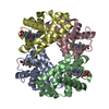 3vreC 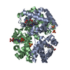 3vrgC 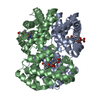 2dn3S S: Starting model for refinement C: citing same article ( |
|---|---|
| Similar structure data |


-Links
 PDBj
PDBj


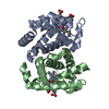
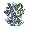
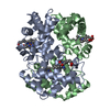
- Assembly
| Deposited unit | 
| ||||||||
|---|---|---|---|---|---|---|---|---|---|
| 1 | 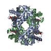
| ||||||||
| Unit cell |
| ||||||||
| Components on special symmetry positions |
|
-Components
| #1: Protein | Mass: 15540.505 Da / Num. of mol.: 1 Source method: isolated from a genetically manipulated source Source: (gene. exp.) Mammuthus primigenius (woolly mammoth) / Strain: SP1349 (KIA27805) / Gene: HBA-T2 / Plasmid: pHE2 / Production host:  Escherichia coli (E. coli) / Strain (production host): JM109 / References: UniProt: D3U1H8 Escherichia coli (E. coli) / Strain (production host): JM109 / References: UniProt: D3U1H8 | ||||
|---|---|---|---|---|---|
| #2: Protein | Mass: 16178.507 Da / Num. of mol.: 1 Source method: isolated from a genetically manipulated source Source: (gene. exp.) Mammuthus primigenius (woolly mammoth) / Strain: SP1349 (KIA27805) / Gene: HBB/D / Plasmid: pHE2 / Production host: Escherichia coli (E. coli) / Strain (production host): JM109 / References: UniProt: D3U1H9 | ||||
| #3: Chemical | Heme B  Mass: 616.487 Da / Num. of mol.: 2 / Source method: obtained synthetically / Formula: C34H32FeN4O4 Mass: 616.487 Da / Num. of mol.: 2 / Source method: obtained synthetically / Formula: C34H32FeN4O4#4: Chemical | Carbon monoxide  Mass: 28.010 Da / Num. of mol.: 2 / Source method: obtained synthetically / Formula: CO Mass: 28.010 Da / Num. of mol.: 2 / Source method: obtained synthetically / Formula: CO#5: Water | ChemComp-HOH / | Water Mass: 18.015 Da / Num. of mol.: 243 / Source method: isolated from a natural source / Formula: H2O Mass: 18.015 Da / Num. of mol.: 243 / Source method: isolated from a natural source / Formula: H2O |
-Experimental details
-Experiment
| Experiment | Method: X-RAY DIFFRACTION / Number of used crystals: 1 |
|---|
- Sample preparation
| Crystal | Density Matthews: 2.68 Å3/Da / Density % sol: 54.07 % |
|---|---|
| Crystal grow | Temperature: 293 K / Method: batch / pH: 7.5 Details: Sodium/potassium phosphate, pH 7.5, BATCH, temperature 293K, temperature 293.0K |
-Data collection
| Diffraction | Mean temperature: 95 K |
|---|---|
| Diffraction source | Source: SYNCHROTRON / Site: Photon Factory  / Beamline: BL-17A / Wavelength: 0.98 Å / Beamline: BL-17A / Wavelength: 0.98 Å |
| Detector | Type: ADSC QUANTUM 270 / Detector: CCD / Date: Mar 17, 2010 / Details: Si(111) |
| Radiation | Monochromator: Si(111) / Protocol: SINGLE WAVELENGTH / Monochromatic (M) / Laue (L): M / Scattering type: x-ray |
| Radiation wavelength | Wavelength: 0.98 Å / Relative weight: 1 |
| Reflection | Resolution: 1.55→50 Å / Num. obs: 47308 / % possible obs: 97.4 % / Observed criterion σ(F): 0 / Observed criterion σ(I): 0 / Redundancy: 2.6 % / Rmerge(I) obs: 0.05 |
| Reflection shell | Resolution: 1.55→1.58 Å / Rmerge(I) obs: 0.187 / % possible all: 83.2 |
- Processing
| Software |
| ||||||||||||||||||||||||||||||||||||||||||||||||||||||||||||||||||||||||||||||||||||||||||||||||||||||||||||||||||||||||||||||||||||||||||||||||||||||||||||||||||||||||||
|---|---|---|---|---|---|---|---|---|---|---|---|---|---|---|---|---|---|---|---|---|---|---|---|---|---|---|---|---|---|---|---|---|---|---|---|---|---|---|---|---|---|---|---|---|---|---|---|---|---|---|---|---|---|---|---|---|---|---|---|---|---|---|---|---|---|---|---|---|---|---|---|---|---|---|---|---|---|---|---|---|---|---|---|---|---|---|---|---|---|---|---|---|---|---|---|---|---|---|---|---|---|---|---|---|---|---|---|---|---|---|---|---|---|---|---|---|---|---|---|---|---|---|---|---|---|---|---|---|---|---|---|---|---|---|---|---|---|---|---|---|---|---|---|---|---|---|---|---|---|---|---|---|---|---|---|---|---|---|---|---|---|---|---|---|---|---|---|---|---|---|---|
| Refinement | Method to determine structure: MOLECULAR REPLACEMENT Starting model: PDB ENTRY 2DN3 Resolution: 1.55→20 Å / Cor.coef. Fo:Fc: 0.961 / Cor.coef. Fo:Fc free: 0.946 / SU B: 1.171 / SU ML: 0.044 / Cross valid method: THROUGHOUT / σ(F): 0 / ESU R: 0.074 / ESU R Free: 0.075 / Stereochemistry target values: MAXIMUM LIKELIHOOD
| ||||||||||||||||||||||||||||||||||||||||||||||||||||||||||||||||||||||||||||||||||||||||||||||||||||||||||||||||||||||||||||||||||||||||||||||||||||||||||||||||||||||||||
| Solvent computation | Ion probe radii: 0.8 Å / Shrinkage radii: 0.8 Å / VDW probe radii: 1.4 Å / Solvent model: BABINET MODEL WITH MASK | ||||||||||||||||||||||||||||||||||||||||||||||||||||||||||||||||||||||||||||||||||||||||||||||||||||||||||||||||||||||||||||||||||||||||||||||||||||||||||||||||||||||||||
| Displacement parameters | Biso mean: 23.417 Å2
| ||||||||||||||||||||||||||||||||||||||||||||||||||||||||||||||||||||||||||||||||||||||||||||||||||||||||||||||||||||||||||||||||||||||||||||||||||||||||||||||||||||||||||
| Refinement step | Cycle: LAST / Resolution: 1.55→20 Å
| ||||||||||||||||||||||||||||||||||||||||||||||||||||||||||||||||||||||||||||||||||||||||||||||||||||||||||||||||||||||||||||||||||||||||||||||||||||||||||||||||||||||||||
| Refine LS restraints |
| ||||||||||||||||||||||||||||||||||||||||||||||||||||||||||||||||||||||||||||||||||||||||||||||||||||||||||||||||||||||||||||||||||||||||||||||||||||||||||||||||||||||||||
| LS refinement shell | Resolution: 1.551→1.591 Å / Total num. of bins used: 20
|
