 Movie
Movie Controller


+ Open data

- Basic information
| Entry | Database: PDB / ID: 3vop | ||||||
|---|---|---|---|---|---|---|---|
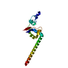
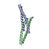
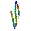
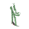
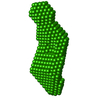
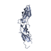
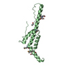
| Title | Structure of Vaccinia virus A27 | ||||||
 Components Components | Protein A27 | ||||||
 Keywords Keywords |  VIRAL PROTEIN / Vaccinia virus / virus fusion protein / virus envelope protein / coiled-coil protein VIRAL PROTEIN / Vaccinia virus / virus fusion protein / virus envelope protein / coiled-coil protein | ||||||
| Function / homology |  Function and homology information Function and homology information | ||||||
| Biological species |   Vaccinia virus Vaccinia virus | ||||||
| Method | X-RAY DIFFRACTION / SYNCHROTRON / MAD / Resolution: 2.2 Å | ||||||
 Authors Authors | Chang, T.H. / Ko, T.P. / Hsieh, F.L. / Wang, A.H.J. | ||||||
 Citation Citation | Journal: Plos Pathog. / Year: 2013 Title: Crystal structure of vaccinia viral A27 protein reveals a novel structure critical for its function and complex formation with A26 protein. Authors: Chang, T.H. / Chang, S.J. / Hsieh, F.L. / Ko, T.P. / Lin, C.T. / Ho, M.R. / Wang, I. / Hsu, S.T. / Guo, R.T. / Chang, W. / Wang, A.H. | ||||||
| History |
|
- Structure visualization
| Structure viewer | Molecule: MolmilJmol/JSmol |
|---|
- Downloads & links
-Download
| PDBx/mmCIF format | 3vop.cif.gz | 70.2 KB | Display | PDBx/mmCIF format |
|---|---|---|---|---|
| PDB format | pdb3vop.ent.gz | 54.5 KB | Display | PDB format |
| PDBx/mmJSON format | 3vop.json.gz | Tree view | PDBx/mmJSON format | |
| Others |  Other downloads Other downloads |
-Validation report
| Arichive directory | https://data.pdbj.org/pub/pdb/validation_reports/vo/3vopftp://data.pdbj.org/pub/pdb/validation_reports/vo/3vop | HTTPS FTP |
|---|
-Related structure data
| Similar structure data |
|---|

-Links
 PDBj
PDBj- Assembly
| Deposited unit | 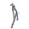
| ||||||||
|---|---|---|---|---|---|---|---|---|---|
| 1 |
| ||||||||
| Unit cell |
|
-Components
| #1: Protein | Mass: 7431.390 Da / Num. of mol.: 3 / Fragment: UNP RESIDUES 21-84 / Mutation: C51A, C52A Source method: isolated from a genetically manipulated source Source: (gene. exp.) Vaccinia virus / Strain: Western Reserve / Gene: A27, A27L, VACWR150 / Plasmid: pET32 / Production host:  Escherichia coli (E. coli) / Strain (production host): BL21 (DE3) / References: UniProt: P11258 Escherichia coli (E. coli) / Strain (production host): BL21 (DE3) / References: UniProt: P11258#2: Chemical | ChemComp-PEG / Diethylene glycol  Mass: 106.120 Da / Num. of mol.: 9 / Source method: obtained synthetically / Formula: C4H10O3 Mass: 106.120 Da / Num. of mol.: 9 / Source method: obtained synthetically / Formula: C4H10O3#3: Water | ChemComp-HOH / | Water Mass: 18.015 Da / Num. of mol.: 130 / Source method: isolated from a natural source / Formula: H2O Mass: 18.015 Da / Num. of mol.: 130 / Source method: isolated from a natural source / Formula: H2O |
|---|
-Experimental details
-Experiment
| Experiment | Method: X-RAY DIFFRACTION / Number of used crystals: 2 |
|---|
- Sample preparation
| Crystal | Density Matthews: 3.17 Å3/Da / Density % sol: 61.23 % / Mosaicity: 0.703 ° |
|---|---|
| Crystal grow | Temperature: 295 K / Method: vapor diffusion / pH: 4.5 Details: 0.1M phosphate-citrate, pH 4.5, 0.2M NaCl, 45-50% PEG 200, vapor diffusion, temperature 295K |
-Data collection
| Diffraction |
| |||||||||||||||||||||||||||||||||||||||||||||||||||||||||||||||||||||||||||||
|---|---|---|---|---|---|---|---|---|---|---|---|---|---|---|---|---|---|---|---|---|---|---|---|---|---|---|---|---|---|---|---|---|---|---|---|---|---|---|---|---|---|---|---|---|---|---|---|---|---|---|---|---|---|---|---|---|---|---|---|---|---|---|---|---|---|---|---|---|---|---|---|---|---|---|---|---|---|---|
| Diffraction source |
| |||||||||||||||||||||||||||||||||||||||||||||||||||||||||||||||||||||||||||||
| Detector |
| |||||||||||||||||||||||||||||||||||||||||||||||||||||||||||||||||||||||||||||
| Radiation |
| |||||||||||||||||||||||||||||||||||||||||||||||||||||||||||||||||||||||||||||
| Radiation wavelength |
| |||||||||||||||||||||||||||||||||||||||||||||||||||||||||||||||||||||||||||||
| Reflection | Redundancy: 18.1 % / Av σ(I) over netI: 46.18 / Number: 163881 / Rmerge(I) obs: 0.077 / Χ2: 1.16 / D res high: 2.62 Å / D res low: 30 Å / Num. obs: 9074 / % possible obs: 99.9 | |||||||||||||||||||||||||||||||||||||||||||||||||||||||||||||||||||||||||||||
| Diffraction reflection shell |
| |||||||||||||||||||||||||||||||||||||||||||||||||||||||||||||||||||||||||||||
| Reflection | Resolution: 2.2→30 Å / Num. all: 15108 / Num. obs: 15074 / % possible obs: 99.3 % / Observed criterion σ(F): 1 / Observed criterion σ(I): 1 / Redundancy: 9.3 % / Biso Wilson estimate: 51.9 Å2 / Rmerge(I) obs: 0.051 / Χ2: 1.295 / Net I/σ(I): 18 | |||||||||||||||||||||||||||||||||||||||||||||||||||||||||||||||||||||||||||||
| Reflection shell |
|


- Processing
| Software |
| ||||||||||||||||||||||||||||||||||||||||||||||||||||||||||||||||||||||||||||||||||||||||||||||||||||
|---|---|---|---|---|---|---|---|---|---|---|---|---|---|---|---|---|---|---|---|---|---|---|---|---|---|---|---|---|---|---|---|---|---|---|---|---|---|---|---|---|---|---|---|---|---|---|---|---|---|---|---|---|---|---|---|---|---|---|---|---|---|---|---|---|---|---|---|---|---|---|---|---|---|---|---|---|---|---|---|---|---|---|---|---|---|---|---|---|---|---|---|---|---|---|---|---|---|---|---|---|---|
| Refinement | Method to determine structure: MAD / Resolution: 2.2→30 Å / Cor.coef. Fo:Fc: 0.943 / Cor.coef. Fo:Fc free: 0.901 / Occupancy max: 1 / Occupancy min: 1 / SU B: 9.779 / SU ML: 0.118 / Cross valid method: THROUGHOUT / σ(F): 0 / σ(I): 0 / ESU R: 0.17 / ESU R Free: 0.179 / Stereochemistry target values: MAXIMUM LIKELIHOOD Details: HYDROGENS HAVE BEEN ADDED IN THE RIDING POSITIONS U VALUES
| ||||||||||||||||||||||||||||||||||||||||||||||||||||||||||||||||||||||||||||||||||||||||||||||||||||
| Solvent computation | Ion probe radii: 0.8 Å / Shrinkage radii: 0.8 Å / VDW probe radii: 1.4 Å / Solvent model: MASK | ||||||||||||||||||||||||||||||||||||||||||||||||||||||||||||||||||||||||||||||||||||||||||||||||||||
| Displacement parameters | Biso max: 100.28 Å2 / Biso mean: 56.506 Å2 / Biso min: 14.13 Å2
| ||||||||||||||||||||||||||||||||||||||||||||||||||||||||||||||||||||||||||||||||||||||||||||||||||||
| Refinement step | Cycle: LAST / Resolution: 2.2→30 Å
| ||||||||||||||||||||||||||||||||||||||||||||||||||||||||||||||||||||||||||||||||||||||||||||||||||||
| Refine LS restraints |
| ||||||||||||||||||||||||||||||||||||||||||||||||||||||||||||||||||||||||||||||||||||||||||||||||||||
| LS refinement shell | Resolution: 2.2→2.257 Å / Total num. of bins used: 20
| ||||||||||||||||||||||||||||||||||||||||||||||||||||||||||||||||||||||||||||||||||||||||||||||||||||
| Refinement TLS params. | Method: refined / Refine-ID: X-RAY DIFFRACTION
| ||||||||||||||||||||||||||||||||||||||||||||||||||||||||||||||||||||||||||||||||||||||||||||||||||||
| Refinement TLS group |
|
