 Movie
Movie Controller

[English] 日本語
 Yorodumi
- PDB-3vn4: Crystal structure of the exosite-containing fragment of human ADA... -
+ Open data

- Basic information
| Entry | Database: PDB / ID: 3vn4 | |||||||||
|---|---|---|---|---|---|---|---|---|---|---|
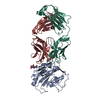
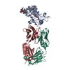
| Title | Crystal structure of the exosite-containing fragment of human ADAMTS13 (P475S mutant) | |||||||||
 Components Components | A disintegrin and metalloproteinase with thrombospondin motifs 13 | |||||||||
 Keywords Keywords |  HYDROLASE HYDROLASE | |||||||||
| Function / homology |  Function and homology informationADAMTS13 endopeptidase / glycoprotein metabolic process / Defective B3GALTL causes PpS / O-glycosylation of TSR domain-containing proteins / response to potassium ion / peptide catabolic process / response to amine / cellular response to interleukin-4 / extracellular matrix organization / cell-matrix adhesion ...ADAMTS13 endopeptidase / glycoprotein metabolic process / Defective B3GALTL causes PpS / O-glycosylation of TSR domain-containing proteins / response to potassium ion / peptide catabolic process / response to amine / cellular response to interleukin-4 / extracellular matrix organization / cell-matrix adhesion / extracellular matrix / integrin-mediated signaling pathway / protein catabolic process / protein processing / response to toxic substance / metalloendopeptidase activity / platelet activation / cellular response to type II interferon / metallopeptidase activity / integrin binding / cellular response to tumor necrosis factor / cellular response to lipopolysaccharide / endoplasmic reticulum lumen / calcium ion binding / cell surface / proteolysis / extracellular space / zinc ion binding Function and homology informationADAMTS13 endopeptidase / glycoprotein metabolic process / Defective B3GALTL causes PpS / O-glycosylation of TSR domain-containing proteins / response to potassium ion / peptide catabolic process / response to amine / cellular response to interleukin-4 / extracellular matrix organization / cell-matrix adhesion ...ADAMTS13 endopeptidase / glycoprotein metabolic process / Defective B3GALTL causes PpS / O-glycosylation of TSR domain-containing proteins / response to potassium ion / peptide catabolic process / response to amine / cellular response to interleukin-4 / extracellular matrix organization / cell-matrix adhesion / extracellular matrix / integrin-mediated signaling pathway / protein catabolic process / protein processing / response to toxic substance / metalloendopeptidase activity / platelet activation / cellular response to type II interferon / metallopeptidase activity / integrin binding / cellular response to tumor necrosis factor / cellular response to lipopolysaccharide / endoplasmic reticulum lumen / calcium ion binding / cell surface / proteolysis / extracellular space / zinc ion bindingSimilarity search - Function | |||||||||
| Biological species |  Homo sapiens (human) Homo sapiens (human) | |||||||||
| Method | X-RAY DIFFRACTION / SYNCHROTRON / MOLECULAR REPLACEMENT / Resolution: 2.8 Å | |||||||||
 Authors Authors | Nakayama, D. / Akiyama, M. / Takeda, S. / Kokame, K. / Takagi, J. / Miyata, T. | |||||||||
 Citation Citation | Journal: To be Published Title: Structural analysis and biochemical studies of ADAMTS13 P475S mutant Authors: Nakayama, D. / Akiyama, M. / Takeda, S. / Kokame, K. / Takagi, J. / Miyata, T. #1: Journal: Proc.Natl.Acad.Sci.USA / Year: 2009 Title: Crystal structures of the noncatalytic domains of ADAMTS13 reveal multiple discontinuous exosites for von Willebrand factor Authors: Akiyama, M. / Takeda, S. / Kokame, K. / Takagi, J. / Miyata, T. #2: Journal: J.Thromb.Haemost. / Year: 2008 Title: ADAMTS13 P475S polymorphism causes a lowered enzymatic activity and urea lability in vitro Authors: Akiyama, M. / Kokame, K. / Miyata, T. | |||||||||
| History |
|
- Structure visualization
| Structure viewer | Molecule: MolmilJmol/JSmol |
|---|
- Downloads & links
-Download
| PDBx/mmCIF format | 3vn4.cif.gz | 166.4 KB | Display | PDBx/mmCIF format |
|---|---|---|---|---|
| PDB format | pdb3vn4.ent.gz | 130.6 KB | Display | PDB format |
| PDBx/mmJSON format | 3vn4.json.gz | Tree view | PDBx/mmJSON format | |
| Others |  Other downloads Other downloads |
-Validation report
| Arichive directory | https://data.pdbj.org/pub/pdb/validation_reports/vn/3vn4ftp://data.pdbj.org/pub/pdb/validation_reports/vn/3vn4 | HTTPS FTP |
|---|
-Related structure data
| Related structure data |  3ghmS S: Starting model for refinement |
|---|---|
| Similar structure data |

-Links
 PDBj
PDBj


- Assembly
| Deposited unit | 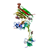
| ||||||||
|---|---|---|---|---|---|---|---|---|---|
| 1 |
| ||||||||
| 2 | 
| ||||||||
| Unit cell |
|
-Components
-Protein / Non-polymers , 2 types, 46 molecules A
| #1: Protein | Mass: 45339.148 Da / Num. of mol.: 1 / Fragment: DTCS DOMAINS, UNP residues 287-685 / Mutation: P475S Source method: isolated from a genetically manipulated source Source: (gene. exp.) Homo sapiens (human) / Gene: ADAMTS13 / Plasmid: PCM-NIDSP / Cell line (production host): HEK293S Gnt1- / Production host:   Cricetulus griseus (Chinese hamster) / References: UniProt: Q76LX8, ADAMTS13 endopeptidase Cricetulus griseus (Chinese hamster) / References: UniProt: Q76LX8, ADAMTS13 endopeptidase |
|---|---|
| #6: Water | ChemComp-HOH / WaterMass: 18.015 Da / Num. of mol.: 45 / Source method: isolated from a natural source / Formula: H2O |
-Sugars , 4 types, 4 molecules 

| #2: Polysaccharide | 2-acetamido-2-deoxy-beta-D-glucopyranose-(1-4)-2-acetamido-2-deoxy-beta-D-glucopyranose / Mass: 424.401 Da / Num. of mol.: 1 Source method: isolated from a genetically manipulated source |
|---|---|
| #3: Polysaccharide | beta-D-glucopyranose-(1-3)-alpha-L-fucopyranose / Mass: 326.297 Da / Num. of mol.: 1 Source method: isolated from a genetically manipulated source |
| #4: Sugar | ChemComp-NAG / N-Acetylglucosamine Type: D-saccharide, beta linking / Mass: 221.208 Da / Num. of mol.: 1 Type: D-saccharide, beta linking / Mass: 221.208 Da / Num. of mol.: 1Source method: isolated from a genetically manipulated source Formula: C8H15NO6 |
| #5: Sugar | ChemComp-BMA / Mannose Type: D-saccharide, beta linking / Mass: 180.156 Da / Num. of mol.: 1 Type: D-saccharide, beta linking / Mass: 180.156 Da / Num. of mol.: 1Source method: isolated from a genetically manipulated source Formula: C6H12O6 |
-Experimental details
-Experiment
| Experiment | Method: X-RAY DIFFRACTION / Number of used crystals: 1 |
|---|
- Sample preparation
| Crystal | Density Matthews: 3.21 Å3/Da / Density % sol: 61.66 % |
|---|---|
| Crystal grow | Temperature: 293 K / Method: vapor diffusion, sitting drop / pH: 6 Details: 20% PEG1500, 0.1M MES, pH 6.0, VAPOR DIFFUSION, SITTING DROP, temperature 293K |
-Data collection
| Diffraction | Mean temperature: 100 K |
|---|---|
| Diffraction source | Source: SYNCHROTRON / Site: SPring-8  / Beamline: BL38B1 / Wavelength: 1 Å / Beamline: BL38B1 / Wavelength: 1 Å |
| Detector | Type: ADSC QUANTUM 315 / Detector: CCD / Date: Feb 21, 2011 |
| Radiation | Monochromator: Fixed exit Si (111) double crystal monochromator Protocol: SINGLE WAVELENGTH / Monochromatic (M) / Laue (L): M / Scattering type: x-ray |
| Radiation wavelength | Wavelength: 1 Å / Relative weight: 1 |
| Reflection | Resolution: 2.8→50 Å / Num. all: 14407 / Num. obs: 14278 / % possible obs: 99.1 % / Observed criterion σ(F): 0 / Observed criterion σ(I): 0 / Redundancy: 3.7 % / Rmerge(I) obs: 0.048 / Net I/σ(I): 20.3 |
| Reflection shell | Resolution: 2.8→2.9 Å / Redundancy: 3.7 % / Rmerge(I) obs: 0.31 / Mean I/σ(I) obs: 6.28 / Num. unique all: 1405 / % possible all: 99 |
- Processing
| Software |
| |||||||||||||||||||||||||||||||||||||||||||||||||||||||||||||||||||||||||||||||||||||
|---|---|---|---|---|---|---|---|---|---|---|---|---|---|---|---|---|---|---|---|---|---|---|---|---|---|---|---|---|---|---|---|---|---|---|---|---|---|---|---|---|---|---|---|---|---|---|---|---|---|---|---|---|---|---|---|---|---|---|---|---|---|---|---|---|---|---|---|---|---|---|---|---|---|---|---|---|---|---|---|---|---|---|---|---|---|---|
| Refinement | Method to determine structure: MOLECULAR REPLACEMENT Starting model: PDB ENTRY 3GHM Resolution: 2.8→30.93 Å / Cor.coef. Fo:Fc: 0.926 / Cor.coef. Fo:Fc free: 0.899 / SU B: 30.293 / SU ML: 0.294 / Cross valid method: THROUGHOUT / σ(F): 0 / σ(I): 0 / ESU R Free: 0.374 / Stereochemistry target values: MAXIMUM LIKELIHOOD / Details: HYDROGENS HAVE BEEN ADDED IN THE RIDING POSITIONS
| |||||||||||||||||||||||||||||||||||||||||||||||||||||||||||||||||||||||||||||||||||||
| Solvent computation | Ion probe radii: 0.8 Å / Shrinkage radii: 0.8 Å / VDW probe radii: 1.4 Å / Solvent model: MASK | |||||||||||||||||||||||||||||||||||||||||||||||||||||||||||||||||||||||||||||||||||||
| Displacement parameters | Biso mean: 62.369 Å2
| |||||||||||||||||||||||||||||||||||||||||||||||||||||||||||||||||||||||||||||||||||||
| Refinement step | Cycle: LAST / Resolution: 2.8→30.93 Å
| |||||||||||||||||||||||||||||||||||||||||||||||||||||||||||||||||||||||||||||||||||||
| Refine LS restraints |
| |||||||||||||||||||||||||||||||||||||||||||||||||||||||||||||||||||||||||||||||||||||
| LS refinement shell | Resolution: 2.8→2.87 Å / Total num. of bins used: 20
| |||||||||||||||||||||||||||||||||||||||||||||||||||||||||||||||||||||||||||||||||||||
| Refinement TLS params. | Method: refined / Refine-ID: X-RAY DIFFRACTION
| |||||||||||||||||||||||||||||||||||||||||||||||||||||||||||||||||||||||||||||||||||||
| Refinement TLS group |
|
