 Movie
Movie Controller

[English] 日本語
 Yorodumi
- PDB-3vmh: Oxygen-bound complex between oxygenase and ferredoxin in carbazol... -
+ Open data

- Basic information
| Entry | Database: PDB / ID: 3vmh | ||||||
|---|---|---|---|---|---|---|---|
| Title | Oxygen-bound complex between oxygenase and ferredoxin in carbazole 1,9a-dioxygenase | ||||||
 Components Components |
| ||||||
 Keywords Keywords |  OXIDOREDUCTASE / Catalytic mechanism / Electron transfer complex / Rieske nonheme iron oxygenase system / Terminal oxygenase / Rieske-type ferredoxin / carbazole 1 / 9a-dioxygenase OXIDOREDUCTASE / Catalytic mechanism / Electron transfer complex / Rieske nonheme iron oxygenase system / Terminal oxygenase / Rieske-type ferredoxin / carbazole 1 / 9a-dioxygenase | ||||||
| Function / homology |  Function and homology information Function and homology information: / carbazole catabolic process / organonitrogen compound catabolic process / ferredoxin hydrogenase activity / dioxygenase activity / 2 iron, 2 sulfur cluster binding / oxidoreductase activity / metal ion bindingSimilarity search - Function | ||||||
| Biological species |  Janthinobacterium (bacteria)Pseudomonas resinovorans (bacteria) Janthinobacterium (bacteria)Pseudomonas resinovorans (bacteria) | ||||||
| Method | X-RAY DIFFRACTION / SYNCHROTRON / MOLECULAR REPLACEMENT / Resolution: 1.85 Å | ||||||
 Authors Authors | Ashikawa, Y. / Nojiri, H. | ||||||
 Citation Citation | Journal: Bmc Struct.Biol. / Year: 2012 Title: Structural insight into the substrate- and dioxygenbinding manner in the catalytic cycle of rieske nonheme iron oxygenase system, carbazole 1,9adioxygenase Authors: Ashikawa, Y. / Fujimoto, Z. / Usami, Y. / Inoue, K. / Noguchi, H. / Yamane, H. / Nojiri, H. | ||||||
| History |
|
- Structure visualization
| Structure viewer | Molecule: MolmilJmol/JSmol |
|---|
- Downloads & links
-Download
| PDBx/mmCIF format | 3vmh.cif.gz | 326.5 KB | Display | PDBx/mmCIF format |
|---|---|---|---|---|
| PDB format | pdb3vmh.ent.gz | 261.4 KB | Display | PDB format |
| PDBx/mmJSON format | 3vmh.json.gz | Tree view | PDBx/mmJSON format | |
| Others |  Other downloads Other downloads |
-Validation report
| Arichive directory | https://data.pdbj.org/pub/pdb/validation_reports/vm/3vmhftp://data.pdbj.org/pub/pdb/validation_reports/vm/3vmh | HTTPS FTP |
|---|
-Related structure data
| Related structure data |  3vmgC  3vmiC 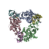 2de5S C: citing same article ( S: Starting model for refinement |
|---|---|
| Similar structure data |


-Links
 PDBj
PDBj


- Assembly
| Deposited unit | 
| ||||||||
|---|---|---|---|---|---|---|---|---|---|
| 1 |
| ||||||||
| Unit cell |
|
-Components
-Protein , 2 types, 6 molecules ABCDEF
| #1: Protein | Mass: 44910.738 Da / Num. of mol.: 3 Source method: isolated from a genetically manipulated source Source: (gene. exp.) Janthinobacterium (bacteria) / Strain: J3 / Gene: carAa / Plasmid: pEJ3AaC / Production host: Escherichia coli (E. coli) / Strain (production host): BL21(DE3) / References: UniProt: Q84II6, EC: 1.14.12.22#2: Protein | / Ferredoxin component of carbazole 1 / 9a-dioxygenaseMass: 12448.059 Da / Num. of mol.: 3 Source method: isolated from a genetically manipulated source Source: (gene. exp.) Pseudomonas resinovorans (bacteria) / Strain: CA10 / Gene: carAc / Plasmid: pECAC1 / Production host: Escherichia coli (E. coli) / Strain (production host): BL21(DE3) / References: UniProt: Q8GI16, EC: 1.14.12.22 |
|---|
-Non-polymers , 4 types, 1242 molecules 


| #3: Chemical |  Mass: 55.845 Da / Num. of mol.: 3 / Source method: obtained synthetically / Formula: Fe Mass: 55.845 Da / Num. of mol.: 3 / Source method: obtained synthetically / Formula: Fe#4: Chemical | ChemComp-FES / Iron–sulfur cluster Mass: 175.820 Da / Num. of mol.: 6 / Source method: obtained synthetically / Formula: Fe2S2 Mass: 175.820 Da / Num. of mol.: 6 / Source method: obtained synthetically / Formula: Fe2S2#5: Chemical | Oxygen Mass: 31.999 Da / Num. of mol.: 3 / Source method: obtained synthetically / Formula: O2 Mass: 31.999 Da / Num. of mol.: 3 / Source method: obtained synthetically / Formula: O2#6: Water | ChemComp-HOH / | WaterMass: 18.015 Da / Num. of mol.: 1230 / Source method: isolated from a natural source / Formula: H2O |
|---|
-Experimental details
-Experiment
| Experiment | Method: X-RAY DIFFRACTION / Number of used crystals: 1 |
|---|
- Sample preparation
| Crystal | Density Matthews: 2.6 Å3/Da / Density % sol: 52.62 % |
|---|---|
| Crystal grow | Temperature: 293 K / Method: vapor diffusion, hanging drop / pH: 5.5 Details: 0.1M ammonium acetate, 12.5% PEG 3350, 0.05M Bis-Tris, pH 5.5, VAPOR DIFFUSION, HANGING DROP, temperature 293K |
-Data collection
| Diffraction | Mean temperature: 100 K |
|---|---|
| Diffraction source | Source: SYNCHROTRON / Site: SPring-8  / Beamline: BL41XU / Wavelength: 1 Å / Beamline: BL41XU / Wavelength: 1 Å |
| Detector | Type: ADSC QUANTUM 315 / Detector: CCD / Date: Jun 28, 2005 |
| Radiation | Monochromator: Rotated-inclined double-crystal monochromator, liquid nitrogen cooling Protocol: SINGLE WAVELENGTH / Monochromatic (M) / Laue (L): M / Scattering type: x-ray |
| Radiation wavelength | Wavelength: 1 Å / Relative weight: 1 |
| Reflection | Resolution: 1.85→50 Å / Num. obs: 150169 / % possible obs: 99.9 % / Biso Wilson estimate: 22.6 Å2 / Rmerge(I) obs: 0.06 |
| Reflection shell | Resolution: 1.85→1.92 Å / Rmerge(I) obs: 0.363 / Num. unique all: 14838 / % possible all: 99.4 |
- Processing
| Software |
| ||||||||||||||||||||||||
|---|---|---|---|---|---|---|---|---|---|---|---|---|---|---|---|---|---|---|---|---|---|---|---|---|---|
| Refinement | Method to determine structure: MOLECULAR REPLACEMENT Starting model: PDB ENTRY 2DE5 Resolution: 1.85→47.61 Å / Rfactor Rfree error: 0.003 / Data cutoff high absF: 2277080.5 / Data cutoff low absF: 0 / Isotropic thermal model: RESTRAINED / Cross valid method: THROUGHOUT / σ(F): 0
| ||||||||||||||||||||||||
| Solvent computation | Solvent model: FLAT MODEL / Bsol: 45.5427 Å2 / ksol: 0.36181 e/Å3 | ||||||||||||||||||||||||
| Displacement parameters | Biso mean: 32 Å2
| ||||||||||||||||||||||||
| Refine analyze |
| ||||||||||||||||||||||||
| Refinement step | Cycle: LAST / Resolution: 1.85→47.61 Å
| ||||||||||||||||||||||||
| Refine LS restraints |
| ||||||||||||||||||||||||
| LS refinement shell | Resolution: 1.85→1.97 Å / Rfactor Rfree error: 0.008 / Total num. of bins used: 6
| ||||||||||||||||||||||||
| Xplor file |
|
