 Movie
Movie Controller

[English] 日本語
 Yorodumi
- PDB-3vm8: Crystal structure of the human APOBEC3C having HIV-1 Vif-binding ... -
+ Open data

- Basic information
| Entry | Database: PDB / ID: 3vm8 | ||||||
|---|---|---|---|---|---|---|---|
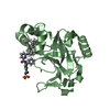
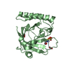
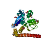
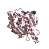
| Title | Crystal structure of the human APOBEC3C having HIV-1 Vif-binding interface | ||||||
 Components Components | Probable DNA dC->dU-editing enzyme APOBEC-3C | ||||||
 Keywords Keywords |  HYDROLASE / APOBEC3C / SIVagm / metal-binding / single domain / Z2 / antiviral defense / host-virus interaction / HIV / HIV-1 Vif / Bet HYDROLASE / APOBEC3C / SIVagm / metal-binding / single domain / Z2 / antiviral defense / host-virus interaction / HIV / HIV-1 Vif / Bet | ||||||
| Function / homology |  Function and homology information Function and homology informationmRNA Editing: C to U Conversion / Formation of the Editosome / cytidine deamination / single-stranded DNA cytosine deaminase / DNA cytosine deamination / cytidine to uridine editing / : / cytidine deaminase activity / clearance of foreign intracellular DNA / negative regulation of single stranded viral RNA replication via double stranded DNA intermediate ...mRNA Editing: C to U Conversion / Formation of the Editosome / cytidine deamination / single-stranded DNA cytosine deaminase / DNA cytosine deamination / cytidine to uridine editing / : / cytidine deaminase activity / clearance of foreign intracellular DNA / negative regulation of single stranded viral RNA replication via double stranded DNA intermediate / : / retrotransposon silencing / negative regulation of viral genome replication / P-body / defense response to virus / innate immune response / RNA binding / zinc ion binding / nucleus / cytoplasmSimilarity search - Function | ||||||
| Biological species |  Homo sapiens (human) Homo sapiens (human) | ||||||
| Method | X-RAY DIFFRACTION / MOLECULAR REPLACEMENT / Resolution: 3 Å | ||||||
 Authors Authors | Kitamura, S. / Suzuki, A. / Watanabe, N. / Iwatani, Y. | ||||||
 Citation Citation | Journal: To be Published Title: Crystal structure of the human APOBEC3C having HIV-1 Vif-binding interface Authors: Kitamura, S. / Ode, H. / Nakashima, M. / Imahashi, M. / Naganawa, Y. / Ibe, S. / Yokomaku, Y. / Watanabe, N. / Suzuki, A. / Sugiura, W. / Iwatani, Y. | ||||||
| History |
|
- Structure visualization
| Structure viewer | Molecule: MolmilJmol/JSmol |
|---|
- Downloads & links
-Download
| PDBx/mmCIF format | 3vm8.cif.gz | 89.5 KB | Display | PDBx/mmCIF format |
|---|---|---|---|---|
| PDB format | pdb3vm8.ent.gz | 68.9 KB | Display | PDB format |
| PDBx/mmJSON format | 3vm8.json.gz | Tree view | PDBx/mmJSON format | |
| Others |  Other downloads Other downloads |
-Validation report
| Arichive directory | https://data.pdbj.org/pub/pdb/validation_reports/vm/3vm8ftp://data.pdbj.org/pub/pdb/validation_reports/vm/3vm8 | HTTPS FTP |
|---|
-Related structure data
| Related structure data | 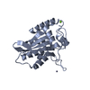 3ir2S S: Starting model for refinement |
|---|---|
| Similar structure data |


-Links
 PDBj
PDBj
- Assembly
| Deposited unit | 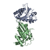
| ||||||||
|---|---|---|---|---|---|---|---|---|---|
| 1 | 
| ||||||||
| 2 | 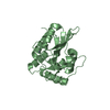
| ||||||||
| Unit cell |
|
-Components
| #1: Protein | Mass: 22851.855 Da / Num. of mol.: 2 Source method: isolated from a genetically manipulated source Source: (gene. exp.) Homo sapiens (human) / Gene: APOBEC3C, APOBEC1L, PBI / Plasmid: pET41a(+) / Production host:  Escherichia coli (E. coli) / Strain (production host): Rosetta2(DE3)pLysS Escherichia coli (E. coli) / Strain (production host): Rosetta2(DE3)pLysSReferences: UniProt: Q9NRW3, Hydrolases; Acting on carbon-nitrogen bonds, other than peptide bonds; In cyclic amidines#2: Chemical |   Mass: 65.409 Da / Num. of mol.: 2 / Source method: obtained synthetically / Formula: Zn Mass: 65.409 Da / Num. of mol.: 2 / Source method: obtained synthetically / Formula: Zn |
|---|
-Experimental details
-Experiment
| Experiment | Method: X-RAY DIFFRACTION / Number of used crystals: 1 |
|---|
- Sample preparation
| Crystal | Density Matthews: 2.57 Å3/Da / Density % sol: 52.05 % / Mosaicity: 0.92 ° |
|---|---|
| Crystal grow | Temperature: 293 K / Method: vapor diffusion, hanging drop / pH: 9 Details: Bicine, PEG 6000, L-Arginine HCl, pH 9.0, vapor diffusion, hanging drop, temperature 293K |
-Data collection
| Diffraction | Mean temperature: 100 K | |||||||||||||||||||||||||||||||||||||||||||||||||||||||||||||||||||||||||||||||||||||||||||||||||||||||||||||||||||||||||||||||||||||||||||||||||||
|---|---|---|---|---|---|---|---|---|---|---|---|---|---|---|---|---|---|---|---|---|---|---|---|---|---|---|---|---|---|---|---|---|---|---|---|---|---|---|---|---|---|---|---|---|---|---|---|---|---|---|---|---|---|---|---|---|---|---|---|---|---|---|---|---|---|---|---|---|---|---|---|---|---|---|---|---|---|---|---|---|---|---|---|---|---|---|---|---|---|---|---|---|---|---|---|---|---|---|---|---|---|---|---|---|---|---|---|---|---|---|---|---|---|---|---|---|---|---|---|---|---|---|---|---|---|---|---|---|---|---|---|---|---|---|---|---|---|---|---|---|---|---|---|---|---|---|---|---|
| Diffraction source | Source: ROTATING ANODE / Type: RIGAKU FR-E SUPERBRIGHT / Wavelength: 1.5418 Å | |||||||||||||||||||||||||||||||||||||||||||||||||||||||||||||||||||||||||||||||||||||||||||||||||||||||||||||||||||||||||||||||||||||||||||||||||||
| Detector | Type: RIGAKU RAXIS VII / Detector: IMAGE PLATE / Date: May 27, 2011 | |||||||||||||||||||||||||||||||||||||||||||||||||||||||||||||||||||||||||||||||||||||||||||||||||||||||||||||||||||||||||||||||||||||||||||||||||||
| Radiation | Monochromator: Confocal Mirror / Protocol: SINGLE WAVELENGTH / Monochromatic (M) / Laue (L): M / Scattering type: x-ray | |||||||||||||||||||||||||||||||||||||||||||||||||||||||||||||||||||||||||||||||||||||||||||||||||||||||||||||||||||||||||||||||||||||||||||||||||||
| Radiation wavelength | Wavelength: 1.5418 Å / Relative weight: 1 | |||||||||||||||||||||||||||||||||||||||||||||||||||||||||||||||||||||||||||||||||||||||||||||||||||||||||||||||||||||||||||||||||||||||||||||||||||
| Reflection | Resolution: 3→1000 Å / Num. all: 9105 / Num. obs: 9105 / % possible obs: 97 % / Observed criterion σ(F): 0 / Observed criterion σ(I): 0 / Redundancy: 10.2 % / Rmerge(I) obs: 0.108 / Χ2: 1.579 / Net I/σ(I): 13.9 | |||||||||||||||||||||||||||||||||||||||||||||||||||||||||||||||||||||||||||||||||||||||||||||||||||||||||||||||||||||||||||||||||||||||||||||||||||
| Reflection shell |
|
- Processing
| Software |
| |||||||||||||||||||||||||||||||||||||||||||||
|---|---|---|---|---|---|---|---|---|---|---|---|---|---|---|---|---|---|---|---|---|---|---|---|---|---|---|---|---|---|---|---|---|---|---|---|---|---|---|---|---|---|---|---|---|---|---|
| Refinement | Method to determine structure: MOLECULAR REPLACEMENT Starting model: PDB ENTRY 3IR2 Resolution: 3→90.43 Å / Cor.coef. Fo:Fc: 0.91 / Cor.coef. Fo:Fc free: 0.833 / WRfactor Rfree: 0.2847 / WRfactor Rwork: 0.2166 / Occupancy max: 1 / Occupancy min: 0.5 / FOM work R set: 0.7549 / SU B: 23.717 / SU ML: 0.441 / SU Rfree: 0.5663 / Cross valid method: THROUGHOUT / σ(F): 0 / ESU R Free: 0.566 / Stereochemistry target values: MAXIMUM LIKELIHOOD
| |||||||||||||||||||||||||||||||||||||||||||||
| Solvent computation | Ion probe radii: 0.8 Å / Shrinkage radii: 0.8 Å / VDW probe radii: 1.2 Å / Solvent model: MASK | |||||||||||||||||||||||||||||||||||||||||||||
| Displacement parameters | Biso max: 126.26 Å2 / Biso mean: 58.0914 Å2 / Biso min: 30.82 Å2
| |||||||||||||||||||||||||||||||||||||||||||||
| Refinement step | Cycle: LAST / Resolution: 3→90.43 Å
| |||||||||||||||||||||||||||||||||||||||||||||
| Refine LS restraints |
| |||||||||||||||||||||||||||||||||||||||||||||
| LS refinement shell | Resolution: 2.998→3.076 Å / Total num. of bins used: 20
|
