 Movie
Movie Controller

[English] 日本語
 Yorodumi
- PDB-3vjr: Crystal structure of Peptidyl-tRNA hydrolase from Escherichia col... -
+ Open data

- Basic information
| Entry | Database: PDB / ID: 3vjr | ||||||
|---|---|---|---|---|---|---|---|
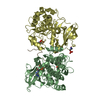
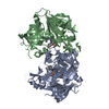
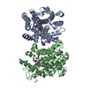
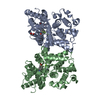
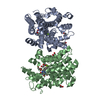
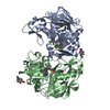
| Title | Crystal structure of Peptidyl-tRNA hydrolase from Escherichia coli in complex with the CCA-acceptor-T[PSI]C domain of tRNA | ||||||
 Components Components |
| ||||||
 Keywords Keywords | hydrolase/RNA /  Peptidyl-tRNA hydrolase / CCA-acceptor-T[PSI]C domain / seven beta-strands form / hydrolase-RNA complex Peptidyl-tRNA hydrolase / CCA-acceptor-T[PSI]C domain / seven beta-strands form / hydrolase-RNA complex | ||||||
| Function / homology |  Function and homology informationpeptidyl-tRNA hydrolase / aminoacyl-tRNA hydrolase activity / protein quality control for misfolded or incompletely synthesized proteins / translation / cytoplasm Function and homology informationpeptidyl-tRNA hydrolase / aminoacyl-tRNA hydrolase activity / protein quality control for misfolded or incompletely synthesized proteins / translation / cytoplasmSimilarity search - Function | ||||||
| Biological species |  Escherichia coli (E. coli) Escherichia coli (E. coli) | ||||||
| Method | X-RAY DIFFRACTION / SYNCHROTRON / MOLECULAR REPLACEMENT / Resolution: 2.4 Å | ||||||
 Authors Authors | Ito, K. / Murakami, R. / Mochizuki, M. / Qi, H. / Shimizu, Y. / Miura, K.I. / Ueda, T. / Uchiumi, T. | ||||||
 Citation Citation | Journal: Nucleic Acids Res. / Year: 2012 Title: Structural basis for the substrate recognition and catalysis of peptidyl-tRNA hydrolase. Authors: Ito, K. / Murakami, R. / Mochizuki, M. / Qi, H. / Shimizu, Y. / Miura, K. / Ueda, T. / Uchiumi, T. | ||||||
| History |
|
- Structure visualization
| Structure viewer | Molecule: MolmilJmol/JSmol |
|---|
- Downloads & links
-Download
| PDBx/mmCIF format | 3vjr.cif.gz | 241.4 KB | Display | PDBx/mmCIF format |
|---|---|---|---|---|
| PDB format | pdb3vjr.ent.gz | 192.1 KB | Display | PDB format |
| PDBx/mmJSON format | 3vjr.json.gz | Tree view | PDBx/mmJSON format | |
| Others |  Other downloads Other downloads |
-Validation report
| Arichive directory | https://data.pdbj.org/pub/pdb/validation_reports/vj/3vjrftp://data.pdbj.org/pub/pdb/validation_reports/vj/3vjr | HTTPS FTP |
|---|
-Related structure data
| Related structure data |  2pthS S: Starting model for refinement |
|---|---|
| Similar structure data |


-Links
 PDBj
PDBj


- Assembly
| Deposited unit | 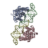
| ||||||||
|---|---|---|---|---|---|---|---|---|---|
| 1 |
| ||||||||
| Unit cell |
|
-Components
| #1: Protein | Alternative ribosome-rescue factor B / PTH Mass: 21395.588 Da / Num. of mol.: 2 Source method: isolated from a genetically manipulated source Source: (gene. exp.) Escherichia coli (E. coli) / Strain: K12 / Gene: pth, b1204, JW1195 / Production host: Escherichia coli (E. coli) / References: UniProt: P0A7D1, peptidyl-tRNA hydrolase#2: RNA chain | Mass: 11532.882 Da / Num. of mol.: 2 / Source method: obtained synthetically / Details: RNA WAS REPEATED BY IN VITRO TRANSCRIPTION #3: Water | ChemComp-HOH / | Water Mass: 18.015 Da / Num. of mol.: 139 / Source method: isolated from a natural source / Formula: H2O Mass: 18.015 Da / Num. of mol.: 139 / Source method: isolated from a natural source / Formula: H2O |
|---|
-Experimental details
-Experiment
| Experiment | Method: X-RAY DIFFRACTION / Number of used crystals: 1 |
|---|
- Sample preparation
| Crystal | Density Matthews: 2.75 Å3/Da / Density % sol: 55.2 % |
|---|---|
| Crystal grow | Temperature: 293 K / Method: vapor diffusion, sitting drop / pH: 5.2 Details: 100mM acetate buffer, 20% (w/v) 1,4-butanediol, 30mM glycyl-glycyl-glycine , pH 5.2, VAPOR DIFFUSION, SITTING DROP, temperature 293K |
-Data collection
| Diffraction | Mean temperature: 100 K |
|---|---|
| Diffraction source | Source: SYNCHROTRON / Site: SPring-8  / Beamline: BL41XU / Wavelength: 1 Å / Beamline: BL41XU / Wavelength: 1 Å |
| Detector | Type: MARMOSAIC 225 mm CCD / Detector: CCD / Date: Jul 22, 2008 |
| Radiation | Monochromator: Si 111 CHANNEL / Protocol: SINGLE WAVELENGTH / Monochromatic (M) / Laue (L): M / Scattering type: x-ray |
| Radiation wavelength | Wavelength: 1 Å / Relative weight: 1 |
| Reflection | Resolution: 2.4→50 Å / Num. all: 27129 / Num. obs: 27129 / % possible obs: 98.1 % / Observed criterion σ(F): 0 / Observed criterion σ(I): 0 / Redundancy: 10.9 % / Rmerge(I) obs: 0.056 / Rsym value: 0.059 / Net I/σ(I): 24.94 |
| Reflection shell | Resolution: 2.4→2.54 Å / Redundancy: 8.1 % / Rmerge(I) obs: 0.187 / Mean I/σ(I) obs: 8.2 / Num. unique all: 4136 / Rsym value: 0.2 / % possible all: 93.7 |
- Processing
| Software |
| |||||||||||||||||||||||||||||||||||||||||||||||||||||||||||||||||||||||||||||||||||||||||||||||||||||||||||||||||||||||||||||
|---|---|---|---|---|---|---|---|---|---|---|---|---|---|---|---|---|---|---|---|---|---|---|---|---|---|---|---|---|---|---|---|---|---|---|---|---|---|---|---|---|---|---|---|---|---|---|---|---|---|---|---|---|---|---|---|---|---|---|---|---|---|---|---|---|---|---|---|---|---|---|---|---|---|---|---|---|---|---|---|---|---|---|---|---|---|---|---|---|---|---|---|---|---|---|---|---|---|---|---|---|---|---|---|---|---|---|---|---|---|---|---|---|---|---|---|---|---|---|---|---|---|---|---|---|---|---|
| Refinement | Method to determine structure: MOLECULAR REPLACEMENT Starting model: 2PTH Resolution: 2.4→50 Å / Cor.coef. Fo:Fc: 0.954 / Cor.coef. Fo:Fc free: 0.931 / SU B: 15.482 / SU ML: 0.164 / Cross valid method: THROUGHOUT / σ(F): 0 / σ(I): 0 / ESU R Free: 0.243 / Stereochemistry target values: MAXIMUM LIKELIHOOD / Details: HYDROGENS HAVE BEEN ADDED IN THE RIDING POSITIONS
| |||||||||||||||||||||||||||||||||||||||||||||||||||||||||||||||||||||||||||||||||||||||||||||||||||||||||||||||||||||||||||||
| Solvent computation | Ion probe radii: 0.8 Å / Shrinkage radii: 0.8 Å / VDW probe radii: 1.4 Å / Solvent model: BABINET MODEL WITH MASK | |||||||||||||||||||||||||||||||||||||||||||||||||||||||||||||||||||||||||||||||||||||||||||||||||||||||||||||||||||||||||||||
| Displacement parameters | Biso mean: 47.222 Å2
| |||||||||||||||||||||||||||||||||||||||||||||||||||||||||||||||||||||||||||||||||||||||||||||||||||||||||||||||||||||||||||||
| Refinement step | Cycle: LAST / Resolution: 2.4→50 Å
| |||||||||||||||||||||||||||||||||||||||||||||||||||||||||||||||||||||||||||||||||||||||||||||||||||||||||||||||||||||||||||||
| Refine LS restraints |
| |||||||||||||||||||||||||||||||||||||||||||||||||||||||||||||||||||||||||||||||||||||||||||||||||||||||||||||||||||||||||||||
| LS refinement shell | Resolution: 2.399→2.462 Å / Total num. of bins used: 20
| |||||||||||||||||||||||||||||||||||||||||||||||||||||||||||||||||||||||||||||||||||||||||||||||||||||||||||||||||||||||||||||
| Refinement TLS params. | Method: refined / Refine-ID: X-RAY DIFFRACTION
| |||||||||||||||||||||||||||||||||||||||||||||||||||||||||||||||||||||||||||||||||||||||||||||||||||||||||||||||||||||||||||||
| Refinement TLS group |
|
