 Movie
Movie Controller


+ Open data

- Basic information
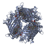
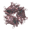
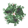
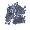
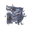
| Entry | Database: PDB / ID: 3vdi | |||||||||
|---|---|---|---|---|---|---|---|---|---|---|
| Title | Structure of the FMO protein from Pelodictyon phaeum | |||||||||
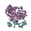
 Components Components | bacteriochlorophyll A protein Bacteriochlorophyll Bacteriochlorophyll | |||||||||
 Keywords Keywords | PHOTOSYNTHESIS / alpha/beta protein / energy transfer | |||||||||
| Function / homology | Bacteriochlorophyll-a Protein / Bacteriochlorophyll A / Clam / Mainly Beta / BACTERIOCHLOROPHYLL A Function and homology information Function and homology information | |||||||||
| Biological species |  Pelodictyon phaeum (bacteria) Pelodictyon phaeum (bacteria) | |||||||||
| Method | X-RAY DIFFRACTION / SYNCHROTRON / MOLECULAR REPLACEMENT / Resolution: 1.99 Å | |||||||||
 Authors Authors | Tronrud, D.E. / Larson, C.R. / Seng, C.O. / Lauman, L. / Matthies, H.J. / Wen, J. / Blankenship, R.E. / Allen, J.P. | |||||||||
 Citation Citation | Journal: Photosynth.Res. / Year: 2012 Title: Reinterpretation of the electron density at the site of the eighth bacteriochlorophyll in the FMO protein from Pelodictyon phaeum. Authors: Tronrud, D.E. / Allen, J.P. #1: Journal: Photosynth.Res. / Year: 2011 Title: The Three-Dimensional Structure of the Fmo Protein from Pelodictyon Phaeum and the Implications for Energy Transfer. Authors: Larson, C.R. / Seng, C.O. / Lauman, L. / Matthies, H.J. / Wen, J. / Blankenship, R.E. / Allen, J.P. | |||||||||
| History |
|
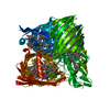
- Structure visualization
| Structure viewer | Molecule: MolmilJmol/JSmol |
|---|
- Downloads & links
-Download
| PDBx/mmCIF format | 3vdi.cif.gz | 183.7 KB | Display | PDBx/mmCIF format |
|---|---|---|---|---|
| PDB format | pdb3vdi.ent.gz | 147.5 KB | Display | PDB format |
| PDBx/mmJSON format | 3vdi.json.gz | Tree view | PDBx/mmJSON format | |
| Others |  Other downloads Other downloads |
-Validation report
| Arichive directory | https://data.pdbj.org/pub/pdb/validation_reports/vd/3vdiftp://data.pdbj.org/pub/pdb/validation_reports/vd/3vdi | HTTPS FTP |
|---|
-Related structure data
| Related structure data | 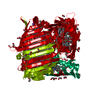 3eojS S: Starting model for refinement |
|---|---|
| Similar structure data |


-Links
 PDBj
PDBj- Assembly
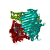
| Deposited unit | 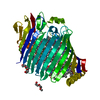
| ||||||||
|---|---|---|---|---|---|---|---|---|---|
| 1 | 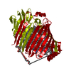
| ||||||||
| Unit cell |
| ||||||||
| Components on special symmetry positions |
|
-Components
| #1: Protein | Bacteriochlorophyll Mass: 40619.746 Da / Num. of mol.: 1 / Source method: isolated from a natural source / Source: (natural) Pelodictyon phaeum (bacteria) | ||||
|---|---|---|---|---|---|
| #2: Chemical | ChemComp-BCL / Bacteriochlorophyll  Mass: 911.504 Da / Num. of mol.: 7 / Source method: obtained synthetically / Formula: C55H74MgN4O6 Mass: 911.504 Da / Num. of mol.: 7 / Source method: obtained synthetically / Formula: C55H74MgN4O6#3: Chemical | ChemComp-PG4 / Polyethylene glycol  Mass: 194.226 Da / Num. of mol.: 4 / Source method: obtained synthetically / Formula: C8H18O5 / Comment: precipitant*YM Mass: 194.226 Da / Num. of mol.: 4 / Source method: obtained synthetically / Formula: C8H18O5 / Comment: precipitant*YM#4: Water | ChemComp-HOH / | Water Mass: 18.015 Da / Num. of mol.: 203 / Source method: isolated from a natural source / Formula: H2O Mass: 18.015 Da / Num. of mol.: 203 / Source method: isolated from a natural source / Formula: H2O |
-Experimental details
-Experiment
| Experiment | Method: X-RAY DIFFRACTION / Number of used crystals: 1 |
|---|
- Sample preparation
| Crystal | Density Matthews: 2.9 Å3/Da / Density % sol: 57.57 % |
|---|---|
| Crystal grow | Temperature: 293 K / Method: vapor diffusion, hanging drop / pH: 7.5 Details: 0.1 mM HEPES, pH 7.5, 16% PEG2000 MME, VAPOR DIFFUSION, HANGING DROP, temperature 293K |
-Data collection
| Diffraction | Mean temperature: 80 K |
|---|---|
| Diffraction source | Source: SYNCHROTRON / Site: NSLS  / Beamline: X29A / Wavelength: 1.0809 / Wavelength: 1.0809 Å / Beamline: X29A / Wavelength: 1.0809 / Wavelength: 1.0809 Å |
| Detector | Type: ADSC QUANTUM 315r / Detector: CCD / Date: Sep 1, 2009 |
| Radiation | Monochromator: horizontal focusing sagittal bend second mono crystal with 4:1 magnification ratio and vertically focusing mirror Protocol: SINGLE WAVELENGTH / Monochromatic (M) / Laue (L): M / Scattering type: x-ray |
| Radiation wavelength | Wavelength: 1.0809 Å / Relative weight: 1 |
| Reflection | Resolution: 1.99→48 Å / Num. all: 29980 / Num. obs: 29980 / % possible obs: 94.5 % / Observed criterion σ(F): 0 / Observed criterion σ(I): 1 / Redundancy: 7.7 % / Biso Wilson estimate: 23.02 Å2 / Rmerge(I) obs: 0.186 |
| Reflection shell | Resolution: 1.99→2.05 Å / Rmerge(I) obs: 0.58 / Mean I/σ(I) obs: 1.2 / % possible all: 94 |
- Processing
| Software |
| ||||||||||||||||||||||||||||||||||||||||||||||||||||||||||||||||||||||||||||||||||||||||||||||||||||||||||||
|---|---|---|---|---|---|---|---|---|---|---|---|---|---|---|---|---|---|---|---|---|---|---|---|---|---|---|---|---|---|---|---|---|---|---|---|---|---|---|---|---|---|---|---|---|---|---|---|---|---|---|---|---|---|---|---|---|---|---|---|---|---|---|---|---|---|---|---|---|---|---|---|---|---|---|---|---|---|---|---|---|---|---|---|---|---|---|---|---|---|---|---|---|---|---|---|---|---|---|---|---|---|---|---|---|---|---|---|---|---|
| Refinement | Method to determine structure: MOLECULAR REPLACEMENT Starting model: PDB ENTRY 3EOJ Resolution: 1.99→40 Å / Cor.coef. Fo:Fc: 0.9465 / Cor.coef. Fo:Fc free: 0.9375 / Cross valid method: THROUGHOUT / σ(F): 0 / Stereochemistry target values: Engh & Huber
| ||||||||||||||||||||||||||||||||||||||||||||||||||||||||||||||||||||||||||||||||||||||||||||||||||||||||||||
| Displacement parameters | Biso mean: 23.4 Å2
| ||||||||||||||||||||||||||||||||||||||||||||||||||||||||||||||||||||||||||||||||||||||||||||||||||||||||||||
| Refine analyze | Luzzati coordinate error obs: 0.174 Å | ||||||||||||||||||||||||||||||||||||||||||||||||||||||||||||||||||||||||||||||||||||||||||||||||||||||||||||
| Refinement step | Cycle: LAST / Resolution: 1.99→40 Å
| ||||||||||||||||||||||||||||||||||||||||||||||||||||||||||||||||||||||||||||||||||||||||||||||||||||||||||||
| Refine LS restraints |
| ||||||||||||||||||||||||||||||||||||||||||||||||||||||||||||||||||||||||||||||||||||||||||||||||||||||||||||
| LS refinement shell | Resolution: 1.99→2.06 Å / Total num. of bins used: 15
| ||||||||||||||||||||||||||||||||||||||||||||||||||||||||||||||||||||||||||||||||||||||||||||||||||||||||||||
| Refinement TLS params. | Method: refined / Origin x: -24.1847 Å / Origin y: 34.0687 Å / Origin z: 24.3031 Å
| ||||||||||||||||||||||||||||||||||||||||||||||||||||||||||||||||||||||||||||||||||||||||||||||||||||||||||||
| Refinement TLS group | Selection details: {A|*} |
