 Movie
Movie Controller

[English] 日本語
 Yorodumi
- PDB-3vcd: Computationally Designed Self-assembling Octahedral Cage protein,... -
+ Open data

- Basic information
| Entry | Database: PDB / ID: 3vcd | ||||||
|---|---|---|---|---|---|---|---|
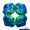
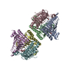
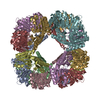
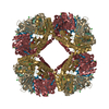
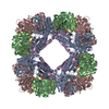
| Title | Computationally Designed Self-assembling Octahedral Cage protein, O333, Crystallized in space group R32 | ||||||
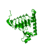
 Components Components | Propanediol utilization polyhedral body protein PduT | ||||||
 Keywords Keywords |  ELECTRON TRANSPORT / self assembling octahedral cage design ELECTRON TRANSPORT / self assembling octahedral cage design | ||||||
| Function / homology |  Function and homology information Function and homology informationpropanediol degradation polyhedral organelle / propanediol catabolic process / 4 iron, 4 sulfur cluster binding / metal ion bindingSimilarity search - Function | ||||||
| Biological species |  Salmonella enterica (bacteria) Salmonella enterica (bacteria) | ||||||
| Method | X-RAY DIFFRACTION / SYNCHROTRON / MOLECULAR REPLACEMENT / molecular replacement / Resolution: 2.35 Å | ||||||
 Authors Authors | Sawaya, M.R. / King, N.P. / Sheffler, W. / Baker, D. / Yeates, T.O. | ||||||
 Citation Citation | Journal: Science / Year: 2012 Title: Computational design of self-assembling protein nanomaterials with atomic level accuracy. Authors: Neil P King / William Sheffler / Michael R Sawaya / Breanna S Vollmar / John P Sumida / Ingemar André / Tamir Gonen / Todd O Yeates / David Baker /  Abstract: We describe a general computational method for designing proteins that self-assemble to a desired symmetric architecture. Protein building blocks are docked together symmetrically to identify ...We describe a general computational method for designing proteins that self-assemble to a desired symmetric architecture. Protein building blocks are docked together symmetrically to identify complementary packing arrangements, and low-energy protein-protein interfaces are then designed between the building blocks in order to drive self-assembly. We used trimeric protein building blocks to design a 24-subunit, 13-nm diameter complex with octahedral symmetry and a 12-subunit, 11-nm diameter complex with tetrahedral symmetry. The designed proteins assembled to the desired oligomeric states in solution, and the crystal structures of the complexes revealed that the resulting materials closely match the design models. The method can be used to design a wide variety of self-assembling protein nanomaterials. | ||||||
| History |
|
- Structure visualization
| Structure viewer | Molecule: MolmilJmol/JSmol |
|---|
- Downloads & links
-Download
| PDBx/mmCIF format | 3vcd.cif.gz | 542 KB | Display | PDBx/mmCIF format |
|---|---|---|---|---|
| PDB format | pdb3vcd.ent.gz | 453.4 KB | Display | PDB format |
| PDBx/mmJSON format | 3vcd.json.gz | Tree view | PDBx/mmJSON format | |
| Others |  Other downloads Other downloads |
-Validation report
| Arichive directory | https://data.pdbj.org/pub/pdb/validation_reports/vc/3vcdftp://data.pdbj.org/pub/pdb/validation_reports/vc/3vcd | HTTPS FTP |
|---|
-Related structure data


-Links
 PDBj
PDBj

- Assembly
| Deposited unit | 
| ||||||||
|---|---|---|---|---|---|---|---|---|---|
| 1 | 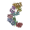
| ||||||||
| Unit cell |
|
-Components
| #1: Protein | Mass: 19964.084 Da / Num. of mol.: 8 Mutation: K15A, C38S, M67L, N148A, N149L, E156S, E160A, K161Y, R167A, V169L Source method: isolated from a genetically manipulated source Source: (gene. exp.) Salmonella enterica (bacteria) / Gene: PduT / Plasmid: pET29b / Production host: Escherichia coli (E. coli) / Strain (production host): BL21(DE3) / References: UniProt: E7V033, UniProt: Q9XDM8*PLUS#2: Chemical | ChemComp-SO4 / Sulfate  Mass: 96.063 Da / Num. of mol.: 26 / Source method: obtained synthetically / Formula: SO4 Mass: 96.063 Da / Num. of mol.: 26 / Source method: obtained synthetically / Formula: SO4#3: Chemical | ChemComp-CL / | Chloride  Mass: 35.453 Da / Num. of mol.: 1 / Source method: obtained synthetically / Formula: Cl Mass: 35.453 Da / Num. of mol.: 1 / Source method: obtained synthetically / Formula: Cl#4: Water | ChemComp-HOH / | Water Mass: 18.015 Da / Num. of mol.: 375 / Source method: isolated from a natural source / Formula: H2O Mass: 18.015 Da / Num. of mol.: 375 / Source method: isolated from a natural source / Formula: H2O |
|---|
-Experimental details
-Experiment
| Experiment | Method: X-RAY DIFFRACTION / Number of used crystals: 1 |
|---|
- Sample preparation
| Crystal | Density Matthews: 3.21 Å3/Da / Density % sol: 61.71 % |
|---|---|
| Crystal grow | Temperature: 298 K / Method: vapor diffusion, hanging drop / pH: 4.5 Details: 0.8M sodium phosphate monobasic, 1.2M potassium phosphate dibasic, 0.1 M sodium acetate, pH 4.5, 10 mM L-proline, crystal soaked in 2M lithium sulfate as cryoprotectant, VAPOR DIFFUSION, ...Details: 0.8M sodium phosphate monobasic, 1.2M potassium phosphate dibasic, 0.1 M sodium acetate, pH 4.5, 10 mM L-proline, crystal soaked in 2M lithium sulfate as cryoprotectant, VAPOR DIFFUSION, HANGING DROP, temperature 298K |
-Data collection
| Diffraction | Mean temperature: 100 K | |||||||||||||||||||||||||||||||||||||||||||||||||||||||||||||||||||||||||||||
|---|---|---|---|---|---|---|---|---|---|---|---|---|---|---|---|---|---|---|---|---|---|---|---|---|---|---|---|---|---|---|---|---|---|---|---|---|---|---|---|---|---|---|---|---|---|---|---|---|---|---|---|---|---|---|---|---|---|---|---|---|---|---|---|---|---|---|---|---|---|---|---|---|---|---|---|---|---|---|
| Diffraction source | Source: SYNCHROTRON / Site: APS / Beamline: 24-ID-C / Wavelength: 0.9794 Å | |||||||||||||||||||||||||||||||||||||||||||||||||||||||||||||||||||||||||||||
| Detector | Type: ADSC QUANTUM 315 / Detector: CCD / Date: Oct 16, 2011 | |||||||||||||||||||||||||||||||||||||||||||||||||||||||||||||||||||||||||||||
| Radiation | Monochromator: Cryo-Cooled Si(111) double crystal / Protocol: SINGLE WAVELENGTH / Monochromatic (M) / Laue (L): M / Scattering type: x-ray | |||||||||||||||||||||||||||||||||||||||||||||||||||||||||||||||||||||||||||||
| Radiation wavelength | Wavelength: 0.9794 Å / Relative weight: 1 | |||||||||||||||||||||||||||||||||||||||||||||||||||||||||||||||||||||||||||||
| Reflection | Resolution: 2.35→100 Å / Num. all: 81734 / Num. obs: 81734 / % possible obs: 95 % / Observed criterion σ(I): -3 / Redundancy: 6.9 % / Biso Wilson estimate: 72.1 Å2 / Rmerge(I) obs: 0.066 / Χ2: 1 / Net I/σ(I): 14.4 | |||||||||||||||||||||||||||||||||||||||||||||||||||||||||||||||||||||||||||||
| Reflection shell |
|
-Phasing
| Phasing | Method: molecular replacement | |||||||||
|---|---|---|---|---|---|---|---|---|---|---|
| Phasing MR | Rfactor: 45.8 / Model details: Phaser MODE: MR_AUTO
|
- Processing
| Software |
| |||||||||||||||||||||||||||||||||||||||||||||||||||||||||||||||||||||||||||||||||||||||||||||||||||||||||||||||||||||||||||||||||||||||||||||||||||||||||||||||||||||||||||||||||||||||||||||||||||||||||||||||||||||||||||||||||
|---|---|---|---|---|---|---|---|---|---|---|---|---|---|---|---|---|---|---|---|---|---|---|---|---|---|---|---|---|---|---|---|---|---|---|---|---|---|---|---|---|---|---|---|---|---|---|---|---|---|---|---|---|---|---|---|---|---|---|---|---|---|---|---|---|---|---|---|---|---|---|---|---|---|---|---|---|---|---|---|---|---|---|---|---|---|---|---|---|---|---|---|---|---|---|---|---|---|---|---|---|---|---|---|---|---|---|---|---|---|---|---|---|---|---|---|---|---|---|---|---|---|---|---|---|---|---|---|---|---|---|---|---|---|---|---|---|---|---|---|---|---|---|---|---|---|---|---|---|---|---|---|---|---|---|---|---|---|---|---|---|---|---|---|---|---|---|---|---|---|---|---|---|---|---|---|---|---|---|---|---|---|---|---|---|---|---|---|---|---|---|---|---|---|---|---|---|---|---|---|---|---|---|---|---|---|---|---|---|---|---|---|---|---|---|---|---|---|---|---|---|---|---|---|---|---|---|
| Refinement | Method to determine structure: MOLECULAR REPLACEMENT / Resolution: 2.35→81.74 Å / Cor.coef. Fo:Fc: 0.9617 / Cor.coef. Fo:Fc free: 0.9446 / Occupancy max: 1 / Occupancy min: 0.5 / SU R Cruickshank DPI: 0.232 / Cross valid method: THROUGHOUT / σ(F): 0 / Stereochemistry target values: Engh & Huber
| |||||||||||||||||||||||||||||||||||||||||||||||||||||||||||||||||||||||||||||||||||||||||||||||||||||||||||||||||||||||||||||||||||||||||||||||||||||||||||||||||||||||||||||||||||||||||||||||||||||||||||||||||||||||||||||||||
| Displacement parameters | Biso max: 179.18 Å2 / Biso mean: 70.1529 Å2 / Biso min: 36.24 Å2
| |||||||||||||||||||||||||||||||||||||||||||||||||||||||||||||||||||||||||||||||||||||||||||||||||||||||||||||||||||||||||||||||||||||||||||||||||||||||||||||||||||||||||||||||||||||||||||||||||||||||||||||||||||||||||||||||||
| Refine analyze | Luzzati coordinate error obs: 0.339 Å | |||||||||||||||||||||||||||||||||||||||||||||||||||||||||||||||||||||||||||||||||||||||||||||||||||||||||||||||||||||||||||||||||||||||||||||||||||||||||||||||||||||||||||||||||||||||||||||||||||||||||||||||||||||||||||||||||
| Refinement step | Cycle: LAST / Resolution: 2.35→81.74 Å
| |||||||||||||||||||||||||||||||||||||||||||||||||||||||||||||||||||||||||||||||||||||||||||||||||||||||||||||||||||||||||||||||||||||||||||||||||||||||||||||||||||||||||||||||||||||||||||||||||||||||||||||||||||||||||||||||||
| Refine LS restraints |
| |||||||||||||||||||||||||||||||||||||||||||||||||||||||||||||||||||||||||||||||||||||||||||||||||||||||||||||||||||||||||||||||||||||||||||||||||||||||||||||||||||||||||||||||||||||||||||||||||||||||||||||||||||||||||||||||||
| LS refinement shell | Resolution: 2.35→2.41 Å / Total num. of bins used: 20
| |||||||||||||||||||||||||||||||||||||||||||||||||||||||||||||||||||||||||||||||||||||||||||||||||||||||||||||||||||||||||||||||||||||||||||||||||||||||||||||||||||||||||||||||||||||||||||||||||||||||||||||||||||||||||||||||||
| Refinement TLS params. | Method: refined / Refine-ID: X-RAY DIFFRACTION
| |||||||||||||||||||||||||||||||||||||||||||||||||||||||||||||||||||||||||||||||||||||||||||||||||||||||||||||||||||||||||||||||||||||||||||||||||||||||||||||||||||||||||||||||||||||||||||||||||||||||||||||||||||||||||||||||||
| Refinement TLS group |
|
