 Movie
Movie Controller


+ Open data

- Basic information
| Entry | Database: PDB / ID: 3v86 | ||||||
|---|---|---|---|---|---|---|---|
| Title | Computational Design of a Protein Crystal | ||||||
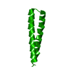
 Components Components | De novo design helix | ||||||
 Keywords Keywords |  DE NOVO PROTEIN / Computational Design of a protein Crystal / Helical Coil / De novo designed helix DE NOVO PROTEIN / Computational Design of a protein Crystal / Helical Coil / De novo designed helix | ||||||
| Method | X-RAY DIFFRACTION / MOLECULAR REPLACEMENT / Resolution: 2.91 Å | ||||||
 Authors Authors | Acharya, R. / North, B. / Saven, J. / DeGrado, W. | ||||||
 Citation Citation | Journal: Proc.Natl.Acad.Sci.USA / Year: 2012 Title: Computational design of a protein crystal. Authors: Lanci, C.J. / Macdermaid, C.M. / Kang, S.G. / Acharya, R. / North, B. / Yang, X. / Qiu, X.J. / Degrado, W.F. / Saven, J.G. | ||||||
| History |
|
- Structure visualization
| Structure viewer | Molecule:  MolmilJmol/JSmol MolmilJmol/JSmol |
|---|
- Downloads & links
-Download
| PDBx/mmCIF format | 3v86.cif.gz | 13.6 KB | Display | PDBx/mmCIF format |
|---|---|---|---|---|
| PDB format | pdb3v86.ent.gz | 8 KB | Display | PDB format |
| PDBx/mmJSON format | 3v86.json.gz | Tree view | PDBx/mmJSON format | |
| Others |  Other downloads Other downloads |
-Validation report
| Arichive directory | https://data.pdbj.org/pub/pdb/validation_reports/v8/3v86ftp://data.pdbj.org/pub/pdb/validation_reports/v8/3v86 | HTTPS FTP |
|---|
-Related structure data
| Related structure data |  4dacC 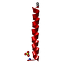 1coiS S: Starting model for refinement C: citing same article ( |
|---|---|
| Similar structure data |


-Links
 PDBj
PDBj
- Assembly
| Deposited unit | 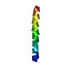
| ||||||||
|---|---|---|---|---|---|---|---|---|---|
| 1 | 
| ||||||||
| Unit cell |
|
-Components
| #1: Protein/peptide | Mass: 3171.639 Da / Num. of mol.: 1 / Source method: obtained synthetically / Details: Chemically Synthesized, De novo design protein |
|---|
-Experimental details
-Experiment
| Experiment | Method: X-RAY DIFFRACTION / Number of used crystals: 1 |
|---|
- Sample preparation
| Crystal | Density Matthews: 2.3 Å3/Da / Density % sol: 46.51 % |
|---|---|
| Crystal grow | Temperature: 298 K / Method: vapor diffusion, hanging drop / pH: 6.5 Details: 10mg/ml peptide solution, well solution, 0.01M Cobalt(II) Chloride hexahydrate, 0.1M MES monohydrate pH 6.5, 1.8M Ammonium sulfate, VAPOR DIFFUSION, HANGING DROP, temperature 298K |
-Data collection
| Diffraction | Mean temperature: 100 K |
|---|---|
| Diffraction source | Source: ROTATING ANODE / Type: RIGAKU / Wavelength: 1.5418 Å |
| Detector | Type: MAR scanner 300 mm plate / Detector: IMAGE PLATE / Date: Jul 13, 2005 / Details: mirrors |
| Radiation | Protocol: SINGLE WAVELENGTH / Monochromatic (M) / Laue (L): M / Scattering type: x-ray |
| Radiation wavelength | Wavelength: 1.5418 Å / Relative weight: 1 |
| Reflection twin | Operator: -h,-k,l / Fraction: 0.505 |
| Reflection | Resolution: 2.89→40.16 Å / Num. all: 741 / Num. obs: 711 / % possible obs: 95.95 % / Observed criterion σ(I): 3 / Redundancy: 8.5 % / Biso Wilson estimate: 51.38 Å2 / Rmerge(I) obs: 0.085 / Net I/σ(I): 26.5 |
| Reflection shell | Resolution: 2.89→3.04 Å / Redundancy: 7.9 % / Rmerge(I) obs: 0.188 / Mean I/σ(I) obs: 11.8 / Num. unique all: 86 / % possible all: 78 |
- Processing
| Software |
| |||||||||||||||||||||||||
|---|---|---|---|---|---|---|---|---|---|---|---|---|---|---|---|---|---|---|---|---|---|---|---|---|---|---|
| Refinement | Method to determine structure: MOLECULAR REPLACEMENT Starting model: poly-alanine model generated from 1COI Resolution: 2.91→40 Å / Isotropic thermal model: 18.8 / Cross valid method: THROUGHOUT / σ(F): 0 / σ(I): 4 / Stereochemistry target values: Engh & Huber Details: REFINED TWIN FRACTION IS 0.505 AND TWIN LAW IS -H, -K, L, USED PROTOCOL IMPLEMENTED IN SHELXL
| |||||||||||||||||||||||||
| Refinement step | Cycle: LAST / Resolution: 2.91→40 Å
| |||||||||||||||||||||||||
| Refine LS restraints |
| |||||||||||||||||||||||||
| LS refinement shell | Resolution: 2.91→3.04 Å
|
