 Movie
Movie Controller

[English] 日本語
 Yorodumi
- PDB-3v3n: Crystal structure of TetX2 T280A: an adaptive mutant in complex w... -
+ Open data

- Basic information
| Entry | Database: PDB / ID: 3v3n | ||||||
|---|---|---|---|---|---|---|---|
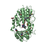
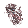
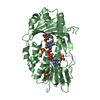
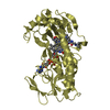
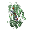
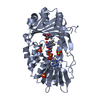
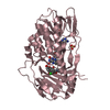
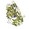
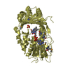
| Title | Crystal structure of TetX2 T280A: an adaptive mutant in complex with minocycline | ||||||
 Components Components | TetX2 protein | ||||||
 Keywords Keywords | OXIDOREDUCTASE/ANTIBIOTIC /  Rossmann Fold / tetracycline degrading monooxygenase / OXIDOREDUCTASE-ANTIBIOTIC complex Rossmann Fold / tetracycline degrading monooxygenase / OXIDOREDUCTASE-ANTIBIOTIC complex | ||||||
| Function / homology |  Function and homology information Function and homology informationtetracycline 11a-monooxygenase / FAD binding / monooxygenase activity / response to antibiotic / cytoplasmSimilarity search - Function | ||||||
| Biological species |  Bacteroides thetaiotaomicron (bacteria) Bacteroides thetaiotaomicron (bacteria) | ||||||
| Method | X-RAY DIFFRACTION / SYNCHROTRON / MOLECULAR REPLACEMENT / molecular replacement / Resolution: 2.703 Å | ||||||
 Authors Authors | Walkiewicz, K. / Shamoo, Y. | ||||||
 Citation Citation | Journal: To be Published Title: Crystal structure of TetX2 T280A: an adaptive mutant in complex with minocycline Authors: Walkiewicz, K. / Shamoo, Y. | ||||||
| History |
|
- Structure visualization
| Structure viewer | Molecule: MolmilJmol/JSmol |
|---|
- Downloads & links
-Download
| PDBx/mmCIF format | 3v3n.cif.gz | 308.6 KB | Display | PDBx/mmCIF format |
|---|---|---|---|---|
| PDB format | pdb3v3n.ent.gz | 248.9 KB | Display | PDB format |
| PDBx/mmJSON format | 3v3n.json.gz | Tree view | PDBx/mmJSON format | |
| Others |  Other downloads Other downloads |
-Validation report
| Arichive directory | https://data.pdbj.org/pub/pdb/validation_reports/v3/3v3nftp://data.pdbj.org/pub/pdb/validation_reports/v3/3v3n | HTTPS FTP |
|---|
-Related structure data
| Related structure data | 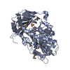 3p9uS S: Starting model for refinement |
|---|---|
| Similar structure data |


-Links
 PDBj
PDBj
- Assembly
| Deposited unit | 
| ||||||||||||||||||||||||||||||||||||||||||||||||||||||||||||||||||||||||||||||||||||||||||||||||
|---|---|---|---|---|---|---|---|---|---|---|---|---|---|---|---|---|---|---|---|---|---|---|---|---|---|---|---|---|---|---|---|---|---|---|---|---|---|---|---|---|---|---|---|---|---|---|---|---|---|---|---|---|---|---|---|---|---|---|---|---|---|---|---|---|---|---|---|---|---|---|---|---|---|---|---|---|---|---|---|---|---|---|---|---|---|---|---|---|---|---|---|---|---|---|---|---|---|
| 1 | 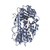
| ||||||||||||||||||||||||||||||||||||||||||||||||||||||||||||||||||||||||||||||||||||||||||||||||
| 2 | 
| ||||||||||||||||||||||||||||||||||||||||||||||||||||||||||||||||||||||||||||||||||||||||||||||||
| 3 | 
| ||||||||||||||||||||||||||||||||||||||||||||||||||||||||||||||||||||||||||||||||||||||||||||||||
| 4 | 
| ||||||||||||||||||||||||||||||||||||||||||||||||||||||||||||||||||||||||||||||||||||||||||||||||
| Unit cell |
| ||||||||||||||||||||||||||||||||||||||||||||||||||||||||||||||||||||||||||||||||||||||||||||||||
| Noncrystallographic symmetry (NCS) | NCS domain:
NCS domain segments: Ens-ID: 1
|