 Movie
Movie Controller

[English] 日本語
 Yorodumi
- PDB-3usv: Structure of the precursor of a thermostable variant of papain at... -
+ Open data

- Basic information
| Entry | Database: PDB / ID: 3usv | ||||||
|---|---|---|---|---|---|---|---|
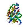
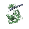
| Title | Structure of the precursor of a thermostable variant of papain at 3.8 A resolution from a crystal soaked at pH 4 | ||||||
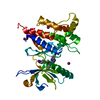
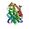
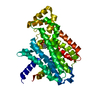
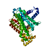
 Components Components | Papain | ||||||
 Keywords Keywords | HYDROLASE / protease | ||||||
| Function / homology |  Function and homology informationpapain / serpin family protein binding / cysteine-type peptidase activity / proteolysis Function and homology informationpapain / serpin family protein binding / cysteine-type peptidase activity / proteolysisSimilarity search - Function | ||||||
| Biological species |  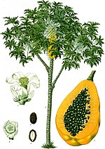 Carica papaya (papaya) Carica papaya (papaya) | ||||||
| Method | X-RAY DIFFRACTION / MOLECULAR REPLACEMENT / Resolution: 3.8 Å | ||||||
 Authors Authors | Roy, S. / Choudhury, D. / Biswas, S. / Dattagupta, J.K. | ||||||
 Citation Citation | Journal: To be Published Title: Crystallographic analysis of pro-papain variant elucidates the structural basis of the step-wise activation mechanism of the zymogen Authors: Roy, S. / Choudhury, D. / Biswas, S. / Dattagupta, J.K. | ||||||
| History |
|
- Structure visualization
| Structure viewer | Molecule: MolmilJmol/JSmol |
|---|
- Downloads & links
-Download
| PDBx/mmCIF format | 3usv.cif.gz | 106.5 KB | Display | PDBx/mmCIF format |
|---|---|---|---|---|
| PDB format | pdb3usv.ent.gz | 67.8 KB | Display | PDB format |
| PDBx/mmJSON format | 3usv.json.gz | Tree view | PDBx/mmJSON format | |
| Others |  Other downloads Other downloads |
-Validation report
| Arichive directory | https://data.pdbj.org/pub/pdb/validation_reports/us/3usvftp://data.pdbj.org/pub/pdb/validation_reports/us/3usv | HTTPS FTP |
|---|
-Related structure data
| Related structure data | 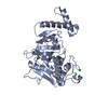 3tnxS S: Starting model for refinement |
|---|---|
| Similar structure data |


-Links
 PDBj
PDBj
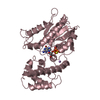
- Assembly
| Deposited unit | 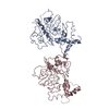
| ||||||||
|---|---|---|---|---|---|---|---|---|---|
| 1 | 
| ||||||||
| 2 | 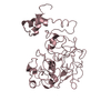
| ||||||||
| Unit cell |
|
-Components
| #1: Protein | / Papaya proteinase I / PPI Mass: 41067.570 Da / Num. of mol.: 2 / Fragment: UNP residues 27-345 / Mutation: C132A, V139S, G143S, K281R Source method: isolated from a genetically manipulated source Source: (gene. exp.) Carica papaya (papaya) / Production host:  Escherichia coli (E. coli) / References: UniProt: P00784, papain Escherichia coli (E. coli) / References: UniProt: P00784, papain |
|---|
-Experimental details
-Experiment
| Experiment | Method: X-RAY DIFFRACTION / Number of used crystals: 1 |
|---|
- Sample preparation
| Crystal | Density Matthews: 2.15 Å3/Da / Density % sol: 42.91 % |
|---|---|
| Crystal grow | Method: vapor diffusion, hanging drop / pH: 4 Details: CS37 of Hampton Research, soaked overnight in NaOAc buffer pH 4.0, VAPOR DIFFUSION, HANGING DROP |
-Data collection
| Diffraction | Mean temperature: 100 K |
|---|---|
| Diffraction source | Source: ROTATING ANODE / Type: Cu FINE FOCUS / Wavelength: 1.54 Å |
| Detector | Type: MAR scanner 345 mm plate / Detector: IMAGE PLATE / Date: May 23, 2011 / Details: Osmic |
| Radiation | Monochromator: Ni / Protocol: SINGLE WAVELENGTH / Monochromatic (M) / Laue (L): M / Scattering type: x-ray |
| Radiation wavelength | Wavelength: 1.54 Å / Relative weight: 1 |
| Reflection | Resolution: 3.8→50 Å / Num. all: 7008 / Num. obs: 6069 / % possible obs: 86.6 % / Observed criterion σ(F): 0 / Observed criterion σ(I): 0 / Rmerge(I) obs: 0.121 / Net I/σ(I): 1.4 |
| Reflection shell | Resolution: 3.8→3.94 Å / % possible all: 88.1 |
- Processing
| Software |
| ||||||||||||||||||||
|---|---|---|---|---|---|---|---|---|---|---|---|---|---|---|---|---|---|---|---|---|---|
| Refinement | Method to determine structure: MOLECULAR REPLACEMENT Starting model: PDB ENTRY 3TNX Resolution: 3.8→29.6 Å / Occupancy max: 1 / Occupancy min: 1 / σ(F): 0 / Stereochemistry target values: Engh & Huber
| ||||||||||||||||||||
| Solvent computation | Bsol: 4294.29 Å2 | ||||||||||||||||||||
| Displacement parameters | Biso max: 60.1 Å2 / Biso mean: 44.4991 Å2 / Biso min: 0.01 Å2
| ||||||||||||||||||||
| Refine analyze | Luzzati coordinate error obs: 0.62 Å | ||||||||||||||||||||
| Refinement step | Cycle: LAST / Resolution: 3.8→29.6 Å
| ||||||||||||||||||||
| Refine LS restraints |
| ||||||||||||||||||||
| LS refinement shell | Resolution: 3.8→4.04 Å / Rfactor Rfree error: 0.044
|
