Mass: 18.015 Da / Num. of mol.: 72 / Source method: isolated from a natural source / Formula: H2O
Sequence details
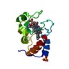
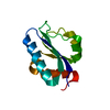
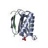
THE CONSTRUCT (RESIDUES 117-213) WAS EXPRESSED WITH A PURIFICATION TAG MGSDKIHHHHHHENLYFQG. THE TAG ...THE CONSTRUCT (RESIDUES 117-213) WAS EXPRESSED WITH A PURIFICATION TAG MGSDKIHHHHHHENLYFQG. THE TAG WAS REMOVED WITH TEV PROTEASE LEAVING ONLY A GLYCINE (0) FOLLOWED BY THE TARGET SEQUENCE. RESIDUE NUMBERING IS BASED ON UNIPROTKB ID P42679.
-
Experimental details
-
Experiment
Experiment
Method: X-RAY DIFFRACTION / Number of used crystals: 1
-
Sample preparation
Crystal
Density Matthews: 2.08 Å3/Da / Density % sol: 40.85 %
Crystal grow
Temperature: 277 K / Method: vapor diffusion, sitting drop / pH: 6 Details: 2.40M (NH4)2SO4, 0.1M MES pH 6.0, NANODROP, VAPOR DIFFUSION, SITTING DROP, temperature 277K
Resolution: 1.5→25.593 Å / Num. obs: 14591 / % possible obs: 96.9 % / Observed criterion σ(I): -3 / Redundancy: 5.02 % / Biso Wilson estimate: 23.592 Å2 / Rmerge(I) obs: 0.047 / Net I/σ(I): 16.87
Reflection shell
Diffraction-ID: 1
Resolution (Å)
Redundancy (%)
Rmerge(I) obs
Mean I/σ(I) obs
Num. measured obs
Num. unique obs
% possible all
1.5-1.54
4.76
0.481
3.29
4927
1036
93.6
1.54-1.58
0.346
4.9
5226
1049
97.8
1.58-1.63
0.263
6.1
5003
996
96.8
1.63-1.68
0.212
7.4
4979
1002
96.2
1.68-1.73
0.154
9.3
4963
991
97.3
1.73-1.79
0.106
12.3
4526
923
97.5
1.79-1.86
0.092
14.1
4431
884
98.2
1.86-1.94
0.071
16.9
4239
872
97.4
1.94-2.02
0.057
19.5
4073
823
97.9
2.02-2.12
0.057
22.7
4359
817
98.4
2.12-2.24
0.058
24
4085
757
97.2
2.24-2.37
0.053
25.1
3915
734
98.3
2.37-2.54
0.05
26.3
3563
669
99
2.54-2.74
0.045
27.8
3273
629
99.1
2.74-3
0.045
28.2
3001
593
98.2
3-3.36
0.043
29.9
2753
537
98
3.36-3.87
0.04
30.7
2411
472
98.3
3.87-4.75
0.038
29.2
1785
385
94.4
4.75-6.71
0.041
28.2
1281
290
91.8
6.71-25.593
0.036
25.5
481
132
71.7
-
Phasing
Phasing
Method: MAD
-
Processing
Software
Name
Version
Classification
NB
MolProbity
3beta29
modelbuilding
PDB_EXTRACT
3.1
dataextraction
SHELX
phasing
SHARP
phasing
XSCALE
December6, 2010
datascaling
BUSTER-TNT
2.10.0
refinement
XDS
datareduction
SHELXD
phasing
BUSTER
2.10.0
refinement
Refinement
Method to determine structure: MAD / Resolution: 1.5→25.593 Å / Cor.coef. Fo:Fc: 0.9632 / Cor.coef. Fo:Fc free: 0.9639 / Occupancy max: 1 / Occupancy min: 0.3 / Cross valid method: THROUGHOUT / σ(F): 0 Details: 1. A MET-INHIBITION PROTOCOL WAS USED FOR SELENOMETHIONINE INCORPORATION DURING PROTEIN EXPRESSION. HOWEVER, THE SE-MET SIDE-CHAIN IS DISORDERED. 2. ATOM RECORD CONTAINS SUM OF TLS AND ...Details: 1. A MET-INHIBITION PROTOCOL WAS USED FOR SELENOMETHIONINE INCORPORATION DURING PROTEIN EXPRESSION. HOWEVER, THE SE-MET SIDE-CHAIN IS DISORDERED. 2. ATOM RECORD CONTAINS SUM OF TLS AND RESIDUAL B FACTORS. ANISOU RECORD CONTAINS SUM OF TLS AND RESIDUAL U FACTORS. 3. SULFATE MODELED IS PRESENT IN CRYSTALLIZATION CONDITIONS. 4. MAD RESTRAINTS (HL COEFFICIENTS) WERE USED DURING REFINEMENT.
In the structure databanks used in Yorodumi, some data are registered as the other names, "COVID-19 virus" and "2019-nCoV". Here are the details of the virus and the list of structure data.
Jan 31, 2019. EMDB accession codes are about to change! (news from PDBe EMDB page)
EMDB accession codes are about to change! (news from PDBe EMDB page)
The allocation of 4 digits for EMDB accession codes will soon come to an end. Whilst these codes will remain in use, new EMDB accession codes will include an additional digit and will expand incrementally as the available range of codes is exhausted. The current 4-digit format prefixed with “EMD-” (i.e. EMD-XXXX) will advance to a 5-digit format (i.e. EMD-XXXXX), and so on. It is currently estimated that the 4-digit codes will be depleted around Spring 2019, at which point the 5-digit format will come into force.
The EM Navigator/Yorodumi systems omit the EMD- prefix.
Related info.:Q: What is EMD? / ID/Accession-code notation in Yorodumi/EM Navigator
Yorodumi is a browser for structure data from EMDB, PDB, SASBDB, etc.
This page is also the successor to EM Navigator detail page, and also detail information page/front-end page for Omokage search.
The word "yorodu" (or yorozu) is an old Japanese word meaning "ten thousand". "mi" (miru) is to see.
Related info.:EMDB / PDB / SASBDB / Comparison of 3 databanks / Yorodumi Search / Aug 31, 2016. New EM Navigator & Yorodumi / Yorodumi Papers / Jmol/JSmol / Function and homology information / Changes in new EM Navigator and Yorodumi
 Movie
Movie Controller

 Yorodumi
 Open data
 Basic information
 Components
 Keywords
 TRANSFERASE /
  Function and homology information
 Authors
 Citation
 Structure visualization
 Downloads & links
 Other downloads


 PDBj
PDBj Assembly
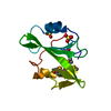


 Mass: 96.063 Da / Num. of mol.: 2 / Source method: obtained synthetically / Formula: SO4
 Mass: 18.015 Da / Num. of mol.: 72 / Source method: isolated from a natural source / Formula: H2O
 Sample preparation
 / Beamline: 8.2.2 / Wavelength: 0.9999,0.9793
 Processing