 Movie
Movie Controller


+ Open data

- Basic information
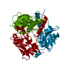
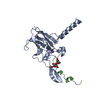
| Entry | Database: PDB / ID: 3unp | ||||||
|---|---|---|---|---|---|---|---|
| Title | Structure of human SUN2 SUN domain | ||||||
 Components Components | SUN domain-containing protein 2 | ||||||
 Keywords Keywords |  TRANSPORT PROTEIN / trimer / nuclear envelope / SUN domain / KASH domain / LINC complex / nuclear migration TRANSPORT PROTEIN / trimer / nuclear envelope / SUN domain / KASH domain / LINC complex / nuclear migration | ||||||
| Function / homology |  Function and homology information Function and homology informationnuclear migration along microfilament / nucleokinesis involved in cell motility in cerebral cortex radial glia guided migration / nuclear matrix anchoring at nuclear membrane / cytoskeleton-nuclear membrane anchor activity / meiotic nuclear membrane microtubule tethering complex / lamin binding / nuclear migration / nuclear inner membrane / centrosome localization / protein-membrane adaptor activity ...nuclear migration along microfilament / nucleokinesis involved in cell motility in cerebral cortex radial glia guided migration / nuclear matrix anchoring at nuclear membrane / cytoskeleton-nuclear membrane anchor activity / meiotic nuclear membrane microtubule tethering complex / lamin binding / nuclear migration / nuclear inner membrane / centrosome localization / protein-membrane adaptor activity / Meiotic synapsis / mitotic spindle organization / meiotic cell cycle / condensed nuclear chromosome / nuclear envelope / microtubule binding / nuclear membrane / chromosome, telomeric region / endosome membrane / positive regulation of cell migration / identical protein bindingSimilarity search - Function | ||||||
| Biological species |  Homo sapiens (human) Homo sapiens (human) | ||||||
| Method | X-RAY DIFFRACTION / SYNCHROTRON / MIR / Resolution: 2.39 Å | ||||||
 Authors Authors | Zhou, Z.C. / Greene, M.I. | ||||||
 Citation Citation | Journal: J.Biol.Chem. / Year: 2012 Title: Structure of Sad1-UNC84 homology (SUN) domain defines features of molecular bridge in nuclear envelope Authors: Zhou, Z.C. / Du, X. / Cai, Z. / Song, X. / Zhang, H. / Mizuno, T. / Suzuki, E. / Yee, M.R. / Berezov, A. / Murali, R. / Wu, S.-L. / Karger, B.L. / Greene, M.I. / Wang, Q. | ||||||
| History |
|
- Structure visualization
| Structure viewer | Molecule: MolmilJmol/JSmol |
|---|
- Downloads & links
-Download
| PDBx/mmCIF format | 3unp.cif.gz | 52.2 KB | Display | PDBx/mmCIF format |
|---|---|---|---|---|
| PDB format | pdb3unp.ent.gz | 38 KB | Display | PDB format |
| PDBx/mmJSON format | 3unp.json.gz | Tree view | PDBx/mmJSON format | |
| Others |  Other downloads Other downloads |
-Validation report
| Arichive directory | https://data.pdbj.org/pub/pdb/validation_reports/un/3unpftp://data.pdbj.org/pub/pdb/validation_reports/un/3unp | HTTPS FTP |
|---|
-Related structure data
| Similar structure data |
|---|


-Links
 PDBj
PDBj
- Assembly
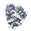
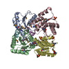
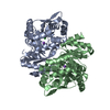
| Deposited unit | 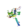
| ||||||||
|---|---|---|---|---|---|---|---|---|---|
| 1 | 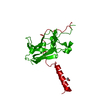
| ||||||||
| Unit cell |
|
-Components
| #1: Protein | Mass: 22885.674 Da / Num. of mol.: 1 / Fragment: SUN domain, residues 520-717 Source method: isolated from a genetically manipulated source Source: (gene. exp.) Homo sapiens (human) / Gene: SUN2 / Production host:  Escherichia coli (E. coli) / References: UniProt: Q9UH99 Escherichia coli (E. coli) / References: UniProt: Q9UH99 |
|---|---|
| #2: Chemical | ChemComp-ACE / Acetyl group  Mass: 44.053 Da / Num. of mol.: 1 / Source method: obtained synthetically / Formula: C2H4O Mass: 44.053 Da / Num. of mol.: 1 / Source method: obtained synthetically / Formula: C2H4O |
| #3: Water | ChemComp-HOH / Water Mass: 18.015 Da / Num. of mol.: 59 / Source method: isolated from a natural source / Formula: H2O Mass: 18.015 Da / Num. of mol.: 59 / Source method: isolated from a natural source / Formula: H2O |
-Experimental details
-Experiment
| Experiment | Method: X-RAY DIFFRACTION / Number of used crystals: 1 |
|---|
- Sample preparation
| Crystal | Density Matthews: 2.59 Å3/Da / Density % sol: 52.53 % |
|---|---|
| Crystal grow | Temperature: 290 K / Method: vapor diffusion, hanging drop / pH: 6.5 Details: 0.1M imidazole, 1.0M sodium acetate, 10 mM YCl3, pH 6.5, VAPOR DIFFUSION, HANGING DROP, temperature 290K |
-Data collection
| Diffraction | Mean temperature: 100 K |
|---|---|
| Diffraction source | Source: SYNCHROTRON / Site: BSRF  / Beamline: 3W1A / Wavelength: 1 Å / Beamline: 3W1A / Wavelength: 1 Å |
| Detector | Type: ADSC QUANTUM 315 / Detector: CCD / Date: May 13, 2008 |
| Radiation | Protocol: SINGLE WAVELENGTH / Monochromatic (M) / Laue (L): M / Scattering type: x-ray |
| Radiation wavelength | Wavelength: 1 Å / Relative weight: 1 |
| Reflection | Resolution: 2.39→50 Å / Num. all: 11047 / Num. obs: 11047 / % possible obs: 100 % / Observed criterion σ(F): 2 / Observed criterion σ(I): 0 |
| Reflection shell | Resolution: 2.85→2.95 Å / % possible all: 100 |
- Processing
| Software |
| ||||||||||||||||||||||||||||||||||||||||||||||||||||||||||||||||||||||||||||||||||||||||||
|---|---|---|---|---|---|---|---|---|---|---|---|---|---|---|---|---|---|---|---|---|---|---|---|---|---|---|---|---|---|---|---|---|---|---|---|---|---|---|---|---|---|---|---|---|---|---|---|---|---|---|---|---|---|---|---|---|---|---|---|---|---|---|---|---|---|---|---|---|---|---|---|---|---|---|---|---|---|---|---|---|---|---|---|---|---|---|---|---|---|---|---|
| Refinement | Method to determine structure: MIR / Resolution: 2.39→39.47 Å / Cor.coef. Fo:Fc: 0.932 / Cor.coef. Fo:Fc free: 0.903 / SU B: 8.048 / SU ML: 0.192 / Cross valid method: THROUGHOUT / σ(F): 2 / ESU R Free: 0.286 / Stereochemistry target values: MAXIMUM LIKELIHOOD
| ||||||||||||||||||||||||||||||||||||||||||||||||||||||||||||||||||||||||||||||||||||||||||
| Solvent computation | Ion probe radii: 0.8 Å / Shrinkage radii: 0.8 Å / VDW probe radii: 1.4 Å / Solvent model: MASK | ||||||||||||||||||||||||||||||||||||||||||||||||||||||||||||||||||||||||||||||||||||||||||
| Displacement parameters | Biso mean: 40.261 Å2
| ||||||||||||||||||||||||||||||||||||||||||||||||||||||||||||||||||||||||||||||||||||||||||
| Refinement step | Cycle: LAST / Resolution: 2.39→39.47 Å
| ||||||||||||||||||||||||||||||||||||||||||||||||||||||||||||||||||||||||||||||||||||||||||
| Refine LS restraints |
| ||||||||||||||||||||||||||||||||||||||||||||||||||||||||||||||||||||||||||||||||||||||||||
| LS refinement shell | Resolution: 2.393→2.454 Å / Total num. of bins used: 20
|
