 Movie
Movie Controller

[English] 日本語
 Yorodumi
- PDB-3uch: Crystal structure of a peptidyl-prolyl cis-trans isomerase E (PPI... -
+ Open data

- Basic information
| Entry | Database: PDB / ID: 3uch | ||||||
|---|---|---|---|---|---|---|---|
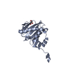
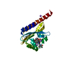
| Title | Crystal structure of a peptidyl-prolyl cis-trans isomerase E (PPIE) from Homo sapiens at 2.50 A resolution | ||||||
 Components Components | Peptidyl-prolyl cis-trans isomerase E | ||||||
 Keywords Keywords |  ISOMERASE / Cyclophilin-like / Structural Genomics / Joint Center for Structural Genomics / JCSG / Protein Structure Initiative / PSI-BIOLOGY / Partnership for T-Cell Biology / TCELL ISOMERASE / Cyclophilin-like / Structural Genomics / Joint Center for Structural Genomics / JCSG / Protein Structure Initiative / PSI-BIOLOGY / Partnership for T-Cell Biology / TCELL | ||||||
| Function / homology |  Function and homology informationpoly(A) binding / U2-type catalytic step 2 spliceosome / cyclosporin A binding / positive regulation of viral genome replication / protein peptidyl-prolyl isomerization / catalytic step 2 spliceosome / mRNA Splicing - Major Pathway / peptidylprolyl isomerase / peptidyl-prolyl cis-trans isomerase activity / Transcription-Coupled Nucleotide Excision Repair (TC-NER) ...poly(A) binding / U2-type catalytic step 2 spliceosome / cyclosporin A binding / positive regulation of viral genome replication / protein peptidyl-prolyl isomerization / catalytic step 2 spliceosome / mRNA Splicing - Major Pathway / peptidylprolyl isomerase / peptidyl-prolyl cis-trans isomerase activity / Transcription-Coupled Nucleotide Excision Repair (TC-NER) / Formation of TC-NER Pre-Incision Complex / mRNA splicing, via spliceosome / Dual incision in TC-NER / Gap-filling DNA repair synthesis and ligation in TC-NER / protein folding / secretory granule lumen / ficolin-1-rich granule lumen / nuclear speck / intracellular membrane-bounded organelle / mRNA binding / Neutrophil degranulation / regulation of DNA-templated transcription / RNA binding / extracellular region / nucleoplasm / nucleus / cytosol / cytoplasm Function and homology informationpoly(A) binding / U2-type catalytic step 2 spliceosome / cyclosporin A binding / positive regulation of viral genome replication / protein peptidyl-prolyl isomerization / catalytic step 2 spliceosome / mRNA Splicing - Major Pathway / peptidylprolyl isomerase / peptidyl-prolyl cis-trans isomerase activity / Transcription-Coupled Nucleotide Excision Repair (TC-NER) ...poly(A) binding / U2-type catalytic step 2 spliceosome / cyclosporin A binding / positive regulation of viral genome replication / protein peptidyl-prolyl isomerization / catalytic step 2 spliceosome / mRNA Splicing - Major Pathway / peptidylprolyl isomerase / peptidyl-prolyl cis-trans isomerase activity / Transcription-Coupled Nucleotide Excision Repair (TC-NER) / Formation of TC-NER Pre-Incision Complex / mRNA splicing, via spliceosome / Dual incision in TC-NER / Gap-filling DNA repair synthesis and ligation in TC-NER / protein folding / secretory granule lumen / ficolin-1-rich granule lumen / nuclear speck / intracellular membrane-bounded organelle / mRNA binding / Neutrophil degranulation / regulation of DNA-templated transcription / RNA binding / extracellular region / nucleoplasm / nucleus / cytosol / cytoplasmSimilarity search - Function | ||||||
| Biological species |  Homo sapiens (human) Homo sapiens (human) | ||||||
| Method | X-RAY DIFFRACTION / SYNCHROTRON / SAD / Resolution: 2.5 Å | ||||||
 Authors Authors | Joint Center for Structural Genomics (JCSG) / Partnership for T-Cell Biology (TCELL) | ||||||
 Citation Citation | Journal: To be published Title: Crystal structure of a Hypotherical Peptidyl-prolyl cis-trans isomerase E (PPIE) from Homo sapiens at 2.50 A resolution Authors: Joint Center for Structural Genomics (JCSG) / Partnership for T-Cell Biology | ||||||
| History |
|
- Structure visualization
| Structure viewer | Molecule: MolmilJmol/JSmol |
|---|
- Downloads & links
-Download
| PDBx/mmCIF format | 3uch.cif.gz | 79.9 KB | Display | PDBx/mmCIF format |
|---|---|---|---|---|
| PDB format | pdb3uch.ent.gz | 63.2 KB | Display | PDB format |
| PDBx/mmJSON format | 3uch.json.gz | Tree view | PDBx/mmJSON format | |
| Others |  Other downloads Other downloads |
-Validation report
| Arichive directory | https://data.pdbj.org/pub/pdb/validation_reports/uc/3uchftp://data.pdbj.org/pub/pdb/validation_reports/uc/3uch | HTTPS FTP |
|---|
-Related structure data
| Similar structure data | |
|---|---|
| Other databases |


-Links
 PDBj
PDBj


- Assembly
| Deposited unit | 
| ||||||||
|---|---|---|---|---|---|---|---|---|---|
| 1 |
| ||||||||
| 2 | 
| ||||||||
| Unit cell |
| ||||||||
| Details | ANALYTICAL SIZE EXCLUSION CHROMATOGRPAHY SUPPORTS THE ASSIGNMENT OF A MONOMER AS A SIGNIFICANT OLIGOMERIZATION STATE IN SOLUTION. HOWEVER, CRYSTAL PACKING ANALYSIS SUGGESTS THAT THE PROTEIN HAS ASSOCIATED INTO TRIMERS THAT ARE PREDICTED TO BE STABLE. |
-Components
| #1: Protein | Mass: 19319.230 Da / Num. of mol.: 1 / Fragment: residues 129-301 Source method: isolated from a genetically manipulated source Source: (gene. exp.) Homo sapiens (human) / Gene: BC008451, CYP33, PPIE / Plasmid: SpeedET / Production host:  Escherichia Coli (E. coli) / Strain (production host): HK100 / References: UniProt: Q9UNP9, peptidylprolyl isomerase Escherichia Coli (E. coli) / Strain (production host): HK100 / References: UniProt: Q9UNP9, peptidylprolyl isomerase |
|---|---|
| #2: Water | ChemComp-HOH / Water Mass: 18.015 Da / Num. of mol.: 49 / Source method: isolated from a natural source / Formula: H2O Mass: 18.015 Da / Num. of mol.: 49 / Source method: isolated from a natural source / Formula: H2O |
| Sequence details | THIS CONSTRUCT WAS EXPRESSED WITH A PURIFICATION TAG MGSDKIHHHHHHENLYFQG. THE TAG WAS REMOVED WITH ...THIS CONSTRUCT WAS EXPRESSED WITH A PURIFICATI |
-Experimental details
-Experiment
| Experiment | Method: X-RAY DIFFRACTION / Number of used crystals: 1 |
|---|
- Sample preparation
| Crystal | Density Matthews: 3.39 Å3/Da / Density % sol: 63.68 % |
|---|---|
| Crystal grow | Temperature: 277 K / Method: vapor diffusion, sitting drop / pH: 7 Details: 20.0% PEG-1000, 0.1M TRIS pH 7.0, NANODROP, VAPOR DIFFUSION, SITTING DROP, temperature 277K |
-Data collection
| Diffraction | Mean temperature: 100 K | ||||||||||||||||||||||||||||||||||||||||||||||||||||||||||||||||||||||||||||||||||||||||||||||||||||||||||||||||||||||||||||||||||||||||||||||||||||||||||||||||||||||||
|---|---|---|---|---|---|---|---|---|---|---|---|---|---|---|---|---|---|---|---|---|---|---|---|---|---|---|---|---|---|---|---|---|---|---|---|---|---|---|---|---|---|---|---|---|---|---|---|---|---|---|---|---|---|---|---|---|---|---|---|---|---|---|---|---|---|---|---|---|---|---|---|---|---|---|---|---|---|---|---|---|---|---|---|---|---|---|---|---|---|---|---|---|---|---|---|---|---|---|---|---|---|---|---|---|---|---|---|---|---|---|---|---|---|---|---|---|---|---|---|---|---|---|---|---|---|---|---|---|---|---|---|---|---|---|---|---|---|---|---|---|---|---|---|---|---|---|---|---|---|---|---|---|---|---|---|---|---|---|---|---|---|---|---|---|---|---|---|---|---|
| Diffraction source | Source: SYNCHROTRON / Site: SSRL  / Beamline: BL11-1 / Wavelength: 0.97894 / Beamline: BL11-1 / Wavelength: 0.97894 | ||||||||||||||||||||||||||||||||||||||||||||||||||||||||||||||||||||||||||||||||||||||||||||||||||||||||||||||||||||||||||||||||||||||||||||||||||||||||||||||||||||||||
| Detector | Type: MARMOSAIC 325 mm CCD / Detector: CCD / Date: Jul 21, 2011 Details: Flat mirror (vertical focusing); single crystal Si(111) bent monochromator (ho rizontal focusing) | ||||||||||||||||||||||||||||||||||||||||||||||||||||||||||||||||||||||||||||||||||||||||||||||||||||||||||||||||||||||||||||||||||||||||||||||||||||||||||||||||||||||||
| Radiation | Monochromator: single crystal Si(111) bent / Protocol: SINGLE WAVELENGTH / Monochromatic (M) / Laue (L): M / Scattering type: x-ray | ||||||||||||||||||||||||||||||||||||||||||||||||||||||||||||||||||||||||||||||||||||||||||||||||||||||||||||||||||||||||||||||||||||||||||||||||||||||||||||||||||||||||
| Radiation wavelength | Wavelength: 0.97894 Å / Relative weight: 1 | ||||||||||||||||||||||||||||||||||||||||||||||||||||||||||||||||||||||||||||||||||||||||||||||||||||||||||||||||||||||||||||||||||||||||||||||||||||||||||||||||||||||||
| Reflection | Resolution: 2.5→29.058 Å / Num. all: 9184 / Num. obs: 9184 / % possible obs: 99.9 % / Redundancy: 23.2 % / Rsym value: 0.149 / Net I/σ(I): 17.2 | ||||||||||||||||||||||||||||||||||||||||||||||||||||||||||||||||||||||||||||||||||||||||||||||||||||||||||||||||||||||||||||||||||||||||||||||||||||||||||||||||||||||||
| Reflection shell | Diffraction-ID: 1
|
-Phasing
| Phasing | Method: SAD |
|---|
- Processing
| Software |
| |||||||||||||||||||||||||||||||||||||||||||||||||||||||||||||||||||||||||||||||||||||
|---|---|---|---|---|---|---|---|---|---|---|---|---|---|---|---|---|---|---|---|---|---|---|---|---|---|---|---|---|---|---|---|---|---|---|---|---|---|---|---|---|---|---|---|---|---|---|---|---|---|---|---|---|---|---|---|---|---|---|---|---|---|---|---|---|---|---|---|---|---|---|---|---|---|---|---|---|---|---|---|---|---|---|---|---|---|---|
| Refinement | Method to determine structure: SAD / Resolution: 2.5→29.058 Å / Cor.coef. Fo:Fc: 0.952 / Cor.coef. Fo:Fc free: 0.939 / Occupancy max: 1 / Occupancy min: 0.5 / SU B: 14.617 / SU ML: 0.169 / Cross valid method: THROUGHOUT / σ(F): 0 / ESU R Free: 0.216 Stereochemistry target values: MAXIMUM LIKELIHOOD WITH PHASES Details: 1. HYDROGENS HAVE BEEN ADDED IN THE RIDING POSITIONS. 2. ATOM RECORD CONTAINS SUM OF TLS AND RESIDUAL B FACTORS. 3. ANISOU RECORD CONTAINS SUM OF TLS AND RESIDUAL U FACTORS. 4. WATERS WERE ...Details: 1. HYDROGENS HAVE BEEN ADDED IN THE RIDING POSITIONS. 2. ATOM RECORD CONTAINS SUM OF TLS AND RESIDUAL B FACTORS. 3. ANISOU RECORD CONTAINS SUM OF TLS AND RESIDUAL U FACTORS. 4. WATERS WERE EXCLUDED FROM AUTOMATIC TLS ASSIGNMENT. 5. A MET-INHIBITION PROTOCOL WAS USED FOR SELENOMETHIONINE INCORPORATION DURING PROTEIN EXPRESSION. THE OCCUPANCY OF THE SE ATOMS IN THE MSE RESIDUES WAS REDUCED TO 0.75 FOR THE REDUCED SCATTERING POWER DUE TO PARTIAL S-MET INCORPORATION.
| |||||||||||||||||||||||||||||||||||||||||||||||||||||||||||||||||||||||||||||||||||||
| Solvent computation | Ion probe radii: 0.8 Å / Shrinkage radii: 0.8 Å / VDW probe radii: 1.4 Å / Solvent model: BABINET MODEL WITH MASK | |||||||||||||||||||||||||||||||||||||||||||||||||||||||||||||||||||||||||||||||||||||
| Displacement parameters | Biso max: 110.33 Å2 / Biso mean: 59.869 Å2 / Biso min: 38.2 Å2 | |||||||||||||||||||||||||||||||||||||||||||||||||||||||||||||||||||||||||||||||||||||
| Refinement step | Cycle: LAST / Resolution: 2.5→29.058 Å
| |||||||||||||||||||||||||||||||||||||||||||||||||||||||||||||||||||||||||||||||||||||
| Refine LS restraints |
| |||||||||||||||||||||||||||||||||||||||||||||||||||||||||||||||||||||||||||||||||||||
| LS refinement shell | Resolution: 2.502→2.567 Å / Total num. of bins used: 20
| |||||||||||||||||||||||||||||||||||||||||||||||||||||||||||||||||||||||||||||||||||||
| Refinement TLS params. | Method: refined / Origin x: 49.585 Å / Origin y: 46.026 Å / Origin z: 17.437 Å
|
