 Movie
Movie Controller

[English] 日本語
 Yorodumi
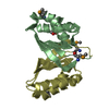
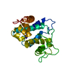
- PDB-3uam: Crystal structure of a chitin binding domain from Burkholderia ps... -
+ Open data

- Basic information
| Entry | Database: PDB / ID: 3uam | ||||||
|---|---|---|---|---|---|---|---|
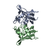
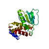
| Title | Crystal structure of a chitin binding domain from Burkholderia pseudomallei | ||||||
 Components Components | Chitin binding domain | ||||||
 Keywords Keywords |  PROTEIN BINDING / SSGCID / Structural Genomics / Seattle Structural Genomics Center for Infectious Disease PROTEIN BINDING / SSGCID / Structural Genomics / Seattle Structural Genomics Center for Infectious Disease | ||||||
| Function / homology | chitin-binding protein cbp21 / Cellulose/chitin-binding protein, N-terminal / Lytic polysaccharide mono-oxygenase, cellulose-degrading / Coagulation Factor XIII; Chain A, domain 1 / Distorted Sandwich / Immunoglobulin E-set / Mainly Beta / NITRATE ION / Chitin binding domain Function and homology information Function and homology information | ||||||
| Biological species |  Burkholderia pseudomallei (bacteria) Burkholderia pseudomallei (bacteria) | ||||||
| Method | X-RAY DIFFRACTION / molecular replacement, molecular replacement / molecular replacement / Resolution: 2 Å | ||||||
 Authors Authors | Seattle Structural Genomics Center for Infectious Disease (SSGCID) | ||||||
 Citation Citation | Journal: to be published Title: Crystal structure of a chitin binding domain from Burkholderia pseudomallei Authors: Seattle Structural Genomics Center for Infectious Disease (SSGCID) / Fox III, D. / Gardberg, A. / Armour, B. / Staker, B. / Stewart, L. | ||||||
| History |
|
- Structure visualization
| Structure viewer | Molecule: MolmilJmol/JSmol |
|---|
- Downloads & links
-Download
| PDBx/mmCIF format | 3uam.cif.gz | 479.9 KB | Display | PDBx/mmCIF format |
|---|---|---|---|---|
| PDB format | pdb3uam.ent.gz | 393.4 KB | Display | PDB format |
| PDBx/mmJSON format | 3uam.json.gz | Tree view | PDBx/mmJSON format | |
| Others |  Other downloads Other downloads |
-Validation report
| Arichive directory | https://data.pdbj.org/pub/pdb/validation_reports/ua/3uamftp://data.pdbj.org/pub/pdb/validation_reports/ua/3uam | HTTPS FTP |
|---|
-Related structure data
| Related structure data | 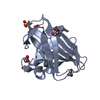 2bemS S: Starting model for refinement |
|---|---|
| Similar structure data | |
| Other databases |

-Links
 PDBj
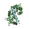
PDBj- Assembly
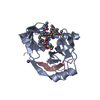
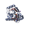
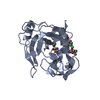
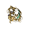
| Deposited unit | 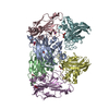
| ||||||||
|---|---|---|---|---|---|---|---|---|---|
| 1 | 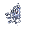
| ||||||||
| 2 | 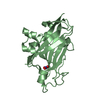
| ||||||||
| 3 | 
| ||||||||
| 4 | 
| ||||||||
| 5 | 
| ||||||||
| 6 | 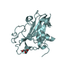
| ||||||||
| Unit cell |
|
-Components
| #1: Protein | Mass: 24191.709 Da / Num. of mol.: 6 Source method: isolated from a genetically manipulated source Source: (gene. exp.) Burkholderia pseudomallei (bacteria) / Strain: 1710b / Gene: BURPS1710b_0114 / Plasmid: AVA0421 / Production host: Escherichia coli (E. coli) / Strain (production host): BL21(DE3) / References: UniProt: Q3JY22#2: Chemical | ChemComp-NO3 / Nitrate  Mass: 62.005 Da / Num. of mol.: 5 / Source method: obtained synthetically / Formula: NO3 Mass: 62.005 Da / Num. of mol.: 5 / Source method: obtained synthetically / Formula: NO3#3: Chemical | Glycerol  Mass: 92.094 Da / Num. of mol.: 3 / Source method: obtained synthetically / Formula: C3H8O3 Mass: 92.094 Da / Num. of mol.: 3 / Source method: obtained synthetically / Formula: C3H8O3#4: Water | ChemComp-HOH / | Water Mass: 18.015 Da / Num. of mol.: 939 / Source method: isolated from a natural source / Formula: H2O Mass: 18.015 Da / Num. of mol.: 939 / Source method: isolated from a natural source / Formula: H2O |
|---|
-Experimental details
-Experiment
| Experiment | Method: X-RAY DIFFRACTION / Number of used crystals: 1 |
|---|
- Sample preparation
| Crystal | Density Matthews: 2.2 Å3/Da / Density % sol: 44.07 % |
|---|---|
| Crystal grow | Temperature: 290 K / pH: 6.77 Details: Internal tracking number 225965. PACT optimization screen F5 well F8. 0.1M Bis Tris propane pH 6.77, 200mM sodium nitrate, 20.54% w/v PEG3500, 20% Ethylene Glycol Cryo. BupsA.17478.a.A1 ...Details: Internal tracking number 225965. PACT optimization screen F5 well F8. 0.1M Bis Tris propane pH 6.77, 200mM sodium nitrate, 20.54% w/v PEG3500, 20% Ethylene Glycol Cryo. BupsA.17478.a.A1 PW31202 21mg/ml, vapor diffusion, sitting drop, temperature 290K |
-Data collection
| Diffraction | Mean temperature: 100 K |
|---|---|
| Diffraction source | Source: ROTATING ANODE / Type: RIGAKU MICROMAX-007 HF / Wavelength: 1.5418 |
| Detector | Type: RIGAKU SATURN 944+ / Detector: CCD / Date: Sep 11, 2011 |
| Radiation | Protocol: SINGLE WAVELENGTH / Monochromatic (M) / Laue (L): M / Scattering type: x-ray |
| Radiation wavelength | Wavelength: 1.5418 Å / Relative weight: 1 |
| Reflection | Resolution: 2→45.013 Å / Num. obs: 82444 / % possible obs: 98.2 % / Observed criterion σ(I): -3 / Biso Wilson estimate: 19.42 Å2 / Rmerge(I) obs: 0.118 / Net I/σ(I): 14.42 |
| Reflection shell | Resolution: 2→2.05 Å / Rmerge(I) obs: 0.437 / Mean I/σ(I) obs: 3.9 / % possible all: 93.5 |
-Phasing
| Phasing | Method: molecular replacement | |||||||||
|---|---|---|---|---|---|---|---|---|---|---|
| Phasing MR | Model details: Phaser MODE: MR_AUTO
|
- Processing
| Software |
| |||||||||||||||||||||||||||||||||||||||||||||||||||||||||||||||||||||||||||||||||||||||||||||||||||||||||||||||||||||||||||||||||||||||||||||||||||||||||||||||||||||||||||||||
|---|---|---|---|---|---|---|---|---|---|---|---|---|---|---|---|---|---|---|---|---|---|---|---|---|---|---|---|---|---|---|---|---|---|---|---|---|---|---|---|---|---|---|---|---|---|---|---|---|---|---|---|---|---|---|---|---|---|---|---|---|---|---|---|---|---|---|---|---|---|---|---|---|---|---|---|---|---|---|---|---|---|---|---|---|---|---|---|---|---|---|---|---|---|---|---|---|---|---|---|---|---|---|---|---|---|---|---|---|---|---|---|---|---|---|---|---|---|---|---|---|---|---|---|---|---|---|---|---|---|---|---|---|---|---|---|---|---|---|---|---|---|---|---|---|---|---|---|---|---|---|---|---|---|---|---|---|---|---|---|---|---|---|---|---|---|---|---|---|---|---|---|---|---|---|---|---|
| Refinement | Method to determine structure: molecular replacement, molecular replacement Starting model: PDB ENTRY 2BEM Resolution: 2→45.013 Å / Cor.coef. Fo:Fc: 0.946 / Cor.coef. Fo:Fc free: 0.918 / SU B: 6.773 / SU ML: 0.101 / Cross valid method: THROUGHOUT / σ(F): 0 / ESU R: 0.188 / ESU R Free: 0.158 / Stereochemistry target values: MAXIMUM LIKELIHOOD Details: U VALUES : WITH TLS ADDED HYDROGENS HAVE BEEN ADDED IN THE RIDING POSITIONS
| |||||||||||||||||||||||||||||||||||||||||||||||||||||||||||||||||||||||||||||||||||||||||||||||||||||||||||||||||||||||||||||||||||||||||||||||||||||||||||||||||||||||||||||||
| Solvent computation | Ion probe radii: 0.8 Å / Shrinkage radii: 0.8 Å / VDW probe radii: 1.2 Å / Solvent model: MASK | |||||||||||||||||||||||||||||||||||||||||||||||||||||||||||||||||||||||||||||||||||||||||||||||||||||||||||||||||||||||||||||||||||||||||||||||||||||||||||||||||||||||||||||||
| Displacement parameters | Biso mean: 14.5 Å2
| |||||||||||||||||||||||||||||||||||||||||||||||||||||||||||||||||||||||||||||||||||||||||||||||||||||||||||||||||||||||||||||||||||||||||||||||||||||||||||||||||||||||||||||||
| Refinement step | Cycle: LAST / Resolution: 2→45.013 Å
| |||||||||||||||||||||||||||||||||||||||||||||||||||||||||||||||||||||||||||||||||||||||||||||||||||||||||||||||||||||||||||||||||||||||||||||||||||||||||||||||||||||||||||||||
| Refine LS restraints |
| |||||||||||||||||||||||||||||||||||||||||||||||||||||||||||||||||||||||||||||||||||||||||||||||||||||||||||||||||||||||||||||||||||||||||||||||||||||||||||||||||||||||||||||||
| LS refinement shell | Resolution: 2→2.05 Å / Total num. of bins used: 20
| |||||||||||||||||||||||||||||||||||||||||||||||||||||||||||||||||||||||||||||||||||||||||||||||||||||||||||||||||||||||||||||||||||||||||||||||||||||||||||||||||||||||||||||||
| Refinement TLS params. | Method: refined / Refine-ID: X-RAY DIFFRACTION
| |||||||||||||||||||||||||||||||||||||||||||||||||||||||||||||||||||||||||||||||||||||||||||||||||||||||||||||||||||||||||||||||||||||||||||||||||||||||||||||||||||||||||||||||
| Refinement TLS group |
|
