 Movie
Movie Controller

[English] 日本語
 Yorodumi
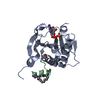
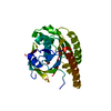
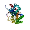
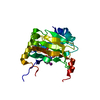
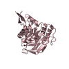
- PDB-3u7x: Crystal structure of the human eIF4E-4EBP1 peptide complex without cap -
+ Open data

- Basic information
| Entry | Database: PDB / ID: 3u7x | |||||||||
|---|---|---|---|---|---|---|---|---|---|---|
| Title | Crystal structure of the human eIF4E-4EBP1 peptide complex without cap | |||||||||
 Components Components |
| |||||||||
 Keywords Keywords |  TRANSLATION / eIF4E / 4EBP1 / mRNA export / structural genomics consortium / SGC TRANSLATION / eIF4E / 4EBP1 / mRNA export / structural genomics consortium / SGC | |||||||||
| Function / homology |  Function and homology information Function and homology informationActivation of the mRNA upon binding of the cap-binding complex and eIFs, and subsequent binding to 43S / eukaryotic initiation factor 4G binding / eukaryotic initiation factor 4E binding / regulation of translation at postsynapse, modulating synaptic transmission / RNA cap binding / chromatoid body / eukaryotic translation initiation factor 4F complex / Z-decay: degradation of maternal mRNAs by zygotically expressed factors / mRNA cap binding / : ...Activation of the mRNA upon binding of the cap-binding complex and eIFs, and subsequent binding to 43S / eukaryotic initiation factor 4G binding / eukaryotic initiation factor 4E binding / regulation of translation at postsynapse, modulating synaptic transmission / RNA cap binding / chromatoid body / eukaryotic translation initiation factor 4F complex / Z-decay: degradation of maternal mRNAs by zygotically expressed factors / mRNA cap binding / : / Deadenylation of mRNA / RNA 7-methylguanosine cap binding / Transport of the SLBP independent Mature mRNA / Transport of the SLBP Dependant Mature mRNA / M-decay: degradation of maternal mRNAs by maternally stored factors / Transport of Mature mRNA Derived from an Intronless Transcript / RISC complex / Ribosomal scanning and start codon recognition / Translation initiation complex formation / stem cell population maintenance / TOR signaling / mTORC1-mediated signalling / GTP hydrolysis and joining of the 60S ribosomal subunit / negative regulation of neuron differentiation / L13a-mediated translational silencing of Ceruloplasmin expression / behavioral fear response / mRNA export from nucleus / translation initiation factor binding / translation repressor activity / negative regulation of translational initiation / translational initiation / translation initiation factor activity / cellular response to dexamethasone stimulus / positive regulation of mitotic cell cycle / P-body / G1/S transition of mitotic cell cycle / neuron differentiation / cytoplasmic ribonucleoprotein granule / ISG15 antiviral mechanism / cytoplasmic stress granule / regulation of translation / postsynapse / DNA-binding transcription factor binding / negative regulation of translation / nuclear speck / glutamatergic synapse / perinuclear region of cytoplasm / enzyme binding / RNA binding / extracellular exosome / nucleus / cytosol / cytoplasmSimilarity search - Function | |||||||||
| Biological species |  Homo sapiens (human) Homo sapiens (human) | |||||||||
| Method | X-RAY DIFFRACTION / SYNCHROTRON / MOLECULAR REPLACEMENT / molecular replacement / Resolution: 2.1 Å | |||||||||
 Authors Authors | Siddiqui, N. / Tempel, W. / Nedyalkova, L. / Wernimont, A.K. / Arrowsmith, C.H. / Edwards, A.M. / Bountra, C. / Weigelt, J. / Borden, K.L.B. / Park, H. / Structural Genomics Consortium (SGC) | |||||||||
 Citation Citation | Journal: J.Mol.Biol. / Year: 2012 Title: Structural Insights into the Allosteric Effects of 4EBP1 on the Eukaryotic Translation Initiation Factor eIF4E. Authors: Siddiqui, N. / Tempel, W. / Nedyalkova, L. / Volpon, L. / Wernimont, A.K. / Osborne, M.J. / Park, H.W. / Borden, K.L. | |||||||||
| History |
|
- Structure visualization
| Structure viewer | Molecule: MolmilJmol/JSmol |
|---|
- Downloads & links
-Download
| PDBx/mmCIF format | 3u7x.cif.gz | 186.2 KB | Display | PDBx/mmCIF format |
|---|---|---|---|---|
| PDB format | pdb3u7x.ent.gz | 147.9 KB | Display | PDB format |
| PDBx/mmJSON format | 3u7x.json.gz | Tree view | PDBx/mmJSON format | |
| Others |  Other downloads Other downloads |
-Validation report
| Arichive directory | https://data.pdbj.org/pub/pdb/validation_reports/u7/3u7xftp://data.pdbj.org/pub/pdb/validation_reports/u7/3u7x | HTTPS FTP |
|---|
-Related structure data
| Related structure data | 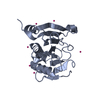 3tf2C 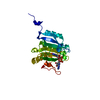 1ipbS S: Starting model for refinement C: citing same article ( |
|---|---|
| Similar structure data |


-Links
 PDBj
PDBj


- Assembly
| Deposited unit | 
| ||||||||||||||||||||||||||||||||||||||||||||||||||||||||||||||||||||
|---|---|---|---|---|---|---|---|---|---|---|---|---|---|---|---|---|---|---|---|---|---|---|---|---|---|---|---|---|---|---|---|---|---|---|---|---|---|---|---|---|---|---|---|---|---|---|---|---|---|---|---|---|---|---|---|---|---|---|---|---|---|---|---|---|---|---|---|---|---|
| 1 | 
| ||||||||||||||||||||||||||||||||||||||||||||||||||||||||||||||||||||
| 2 | 
| ||||||||||||||||||||||||||||||||||||||||||||||||||||||||||||||||||||
| 3 | 
| ||||||||||||||||||||||||||||||||||||||||||||||||||||||||||||||||||||
| Unit cell |
| ||||||||||||||||||||||||||||||||||||||||||||||||||||||||||||||||||||
| Noncrystallographic symmetry (NCS) | NCS domain:
NCS domain segments: Component-ID: 1 / Refine code: 5
NCS ensembles :
|
-Components
| #1: Protein | EIF4E / eIF-4E / eIF4E / eIF-4F 25 kDa subunit / mRNA cap-binding protein Mass: 25130.242 Da / Num. of mol.: 2 Source method: isolated from a genetically manipulated source Source: (gene. exp.) Homo sapiens (human) / Gene: EIF4E, EIF4EL1, EIF4F / Plasmid: pT7 / Production host:  Escherichia coli (E. coli) / Strain (production host): BL21(DE3) / References: UniProt: P06730 Escherichia coli (E. coli) / Strain (production host): BL21(DE3) / References: UniProt: P06730#2: Protein/peptide | Mass: 2357.755 Da / Num. of mol.: 2 / Fragment: UNP residues 46-66 / Source method: obtained synthetically Details: Fmoc solid-phase peptide synthesis (Sheldon Biotechnology Center) Source: (synth.) Homo sapiens (human) / References: UniProt: Q13541#3: Chemical | ChemComp-SO4 / Sulfate  Mass: 96.063 Da / Num. of mol.: 4 / Source method: obtained synthetically / Formula: SO4 Mass: 96.063 Da / Num. of mol.: 4 / Source method: obtained synthetically / Formula: SO4#4: Chemical | ChemComp-UNX /   Num. of mol.: 20 / Source method: obtained synthetically Num. of mol.: 20 / Source method: obtained synthetically#5: Water | ChemComp-HOH / | Water Mass: 18.015 Da / Num. of mol.: 123 / Source method: isolated from a natural source / Formula: H2O Mass: 18.015 Da / Num. of mol.: 123 / Source method: isolated from a natural source / Formula: H2O |
|---|
-Experimental details
-Experiment
| Experiment | Method: X-RAY DIFFRACTION / Number of used crystals: 1 |
|---|
- Sample preparation
| Crystal | Density Matthews: 2.79 Å3/Da / Density % sol: 55.89 % |
|---|---|
| Crystal grow | Temperature: 291 K / Method: vapor diffusion, sitting drop / pH: 7.5 Details: 1:1 of (protein stock: 0.0004 M eif4e, 0.001 M 4epb1, 0.004 M ribavirin, 0.02 HEPES, 0.1 M potassium chloride, 0.001 tcep, ph 7.3):(reservoir: 2 M ammonium sulfate, 2% Peg-400, 0.1 M sodium ...Details: 1:1 of (protein stock: 0.0004 M eif4e, 0.001 M 4epb1, 0.004 M ribavirin, 0.02 HEPES, 0.1 M potassium chloride, 0.001 tcep, ph 7.3):(reservoir: 2 M ammonium sulfate, 2% Peg-400, 0.1 M sodium HEPES), vapor diffusion, sitting drop, temperature 291K |
-Data collection
| Diffraction | Mean temperature: 100 K | |||||||||||||||||||||||||||||||||||||||||||||||||||||||||||||||||||||||||||||||||||||||||||||||||||||||||||||||||||||||||||||||||||||||||||||||||||
|---|---|---|---|---|---|---|---|---|---|---|---|---|---|---|---|---|---|---|---|---|---|---|---|---|---|---|---|---|---|---|---|---|---|---|---|---|---|---|---|---|---|---|---|---|---|---|---|---|---|---|---|---|---|---|---|---|---|---|---|---|---|---|---|---|---|---|---|---|---|---|---|---|---|---|---|---|---|---|---|---|---|---|---|---|---|---|---|---|---|---|---|---|---|---|---|---|---|---|---|---|---|---|---|---|---|---|---|---|---|---|---|---|---|---|---|---|---|---|---|---|---|---|---|---|---|---|---|---|---|---|---|---|---|---|---|---|---|---|---|---|---|---|---|---|---|---|---|---|
| Diffraction source | Source: SYNCHROTRON / Site: APS  / Beamline: 19-ID / Wavelength: 0.97924 Å / Beamline: 19-ID / Wavelength: 0.97924 Å | |||||||||||||||||||||||||||||||||||||||||||||||||||||||||||||||||||||||||||||||||||||||||||||||||||||||||||||||||||||||||||||||||||||||||||||||||||
| Detector | Type: ADSC QUANTUM 315 / Detector: CCD / Date: Aug 18, 2010 | |||||||||||||||||||||||||||||||||||||||||||||||||||||||||||||||||||||||||||||||||||||||||||||||||||||||||||||||||||||||||||||||||||||||||||||||||||
| Radiation | Protocol: SINGLE WAVELENGTH / Monochromatic (M) / Laue (L): M / Scattering type: x-ray | |||||||||||||||||||||||||||||||||||||||||||||||||||||||||||||||||||||||||||||||||||||||||||||||||||||||||||||||||||||||||||||||||||||||||||||||||||
| Radiation wavelength | Wavelength: 0.97924 Å / Relative weight: 1 | |||||||||||||||||||||||||||||||||||||||||||||||||||||||||||||||||||||||||||||||||||||||||||||||||||||||||||||||||||||||||||||||||||||||||||||||||||
| Reflection | Resolution: 2.1→50 Å / Num. obs: 35859 / % possible obs: 99.7 % / Redundancy: 3.6 % / Rmerge(I) obs: 0.089 / Χ2: 1.282 / Net I/σ(I): 7.6 | |||||||||||||||||||||||||||||||||||||||||||||||||||||||||||||||||||||||||||||||||||||||||||||||||||||||||||||||||||||||||||||||||||||||||||||||||||
| Reflection shell |
|
-Phasing
| Phasing | Method: molecular replacement |
|---|
- Processing
| Software |
| |||||||||||||||||||||||||||||||||||||||||||||||||||||||||||||||||||||||||||||||||||||||||||||||||||||||||||||||||||||||||||||||||||||||||||||||||||
|---|---|---|---|---|---|---|---|---|---|---|---|---|---|---|---|---|---|---|---|---|---|---|---|---|---|---|---|---|---|---|---|---|---|---|---|---|---|---|---|---|---|---|---|---|---|---|---|---|---|---|---|---|---|---|---|---|---|---|---|---|---|---|---|---|---|---|---|---|---|---|---|---|---|---|---|---|---|---|---|---|---|---|---|---|---|---|---|---|---|---|---|---|---|---|---|---|---|---|---|---|---|---|---|---|---|---|---|---|---|---|---|---|---|---|---|---|---|---|---|---|---|---|---|---|---|---|---|---|---|---|---|---|---|---|---|---|---|---|---|---|---|---|---|---|---|---|---|---|
| Refinement | Method to determine structure: MOLECULAR REPLACEMENT Starting model: PDB ENTRY 1IPB Resolution: 2.1→30 Å / Cor.coef. Fo:Fc: 0.946 / Cor.coef. Fo:Fc free: 0.928 / WRfactor Rfree: 0.21 / WRfactor Rwork: 0.186 / Occupancy max: 1 / Occupancy min: 0.3 / SU B: 9.957 / SU ML: 0.118 / Cross valid method: THROUGHOUT / σ(F): 0 / ESU R Free: 0.152 / Stereochemistry target values: MAXIMUM LIKELIHOOD Details: HYDROGENS HAVE BEEN ADDED IN THE RIDING POSITIONS U VALUES : WITH TLS ADDED. AMINO ACID RESIDUES 51 THROUGH 58 ARE POORLY DEFINED BY ELECTRON DENSITY. FO-FC DENSITY SUGGESTS AN ALTERNATE ...Details: HYDROGENS HAVE BEEN ADDED IN THE RIDING POSITIONS U VALUES : WITH TLS ADDED. AMINO ACID RESIDUES 51 THROUGH 58 ARE POORLY DEFINED BY ELECTRON DENSITY. FO-FC DENSITY SUGGESTS AN ALTERNATE CONFORMATION OF THIS REGION FOR CHAIN A. AUTOBUSTER, PHENIX, COOT AND THE MOLPROBITY SERVER WERE ALSO USED DURING REFINEMENT OF THE MODEL.
| |||||||||||||||||||||||||||||||||||||||||||||||||||||||||||||||||||||||||||||||||||||||||||||||||||||||||||||||||||||||||||||||||||||||||||||||||||
| Solvent computation | Ion probe radii: 0.8 Å / Shrinkage radii: 0.8 Å / VDW probe radii: 1.4 Å / Solvent model: BABINET MODEL PLUS MASK | |||||||||||||||||||||||||||||||||||||||||||||||||||||||||||||||||||||||||||||||||||||||||||||||||||||||||||||||||||||||||||||||||||||||||||||||||||
| Displacement parameters | Biso max: 114.74 Å2 / Biso mean: 41.169 Å2 / Biso min: 27.09 Å2
| |||||||||||||||||||||||||||||||||||||||||||||||||||||||||||||||||||||||||||||||||||||||||||||||||||||||||||||||||||||||||||||||||||||||||||||||||||
| Refinement step | Cycle: LAST / Resolution: 2.1→30 Å
| |||||||||||||||||||||||||||||||||||||||||||||||||||||||||||||||||||||||||||||||||||||||||||||||||||||||||||||||||||||||||||||||||||||||||||||||||||
| Refine LS restraints |
| |||||||||||||||||||||||||||||||||||||||||||||||||||||||||||||||||||||||||||||||||||||||||||||||||||||||||||||||||||||||||||||||||||||||||||||||||||
| Refine LS restraints NCS | Dom-ID: 1 / Refine-ID: X-RAY DIFFRACTION
| |||||||||||||||||||||||||||||||||||||||||||||||||||||||||||||||||||||||||||||||||||||||||||||||||||||||||||||||||||||||||||||||||||||||||||||||||||
| LS refinement shell | Refine-ID: X-RAY DIFFRACTION / Total num. of bins used: 20
| |||||||||||||||||||||||||||||||||||||||||||||||||||||||||||||||||||||||||||||||||||||||||||||||||||||||||||||||||||||||||||||||||||||||||||||||||||
| Refinement TLS params. | Method: refined / Refine-ID: X-RAY DIFFRACTION
| |||||||||||||||||||||||||||||||||||||||||||||||||||||||||||||||||||||||||||||||||||||||||||||||||||||||||||||||||||||||||||||||||||||||||||||||||||
| Refinement TLS group |
|
