[English] 日本語
Yorodumi
- PDB-3u50: Crystal Structure of the Tetrahymena telomerase processivity fact... -

+
Open data


ID or keywords:

Loading...

-
Basic information

Entry
Database: PDB / ID: 3u50
Title
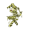
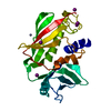
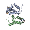
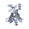
Crystal Structure of the Tetrahymena telomerase processivity factor Teb1 OB-C
ComponentsTelomerase-associated protein 82
KeywordsDNA BINDING PROTEIN / tetrahymena / telomerase / Teb1 / processivity factor
Function / homology
Function and homology information


single-stranded telomeric DNA binding / telomerase holoenzyme complex / telomere maintenance via telomerase / chromosome, telomeric region / zinc ion binding
Similarity search - Function
: / Replication factor A, C-terminal / Replication factor-A C terminal domain / N-terminal domain of TfIIb - #10 / N-terminal domain of TfIIb / Single Sheet / Nucleic acid-binding proteins / OB fold (Dihydrolipoamide Acetyltransferase, E2P) / Nucleic acid-binding, OB-fold / Beta Barrel / Mainly Beta
Similarity search - Domain/homology
Telomeric repeat-binding subunit 1
Similarity search - Component
Biological speciesTetrahymena thermophila (eukaryote)
MethodX-RAY DIFFRACTION / SYNCHROTRON / SAD / Resolution: 2.5 Å
AuthorsZeng, Z. / Huang, J. / Yang, Y. / Lei, M.
CitationJournal: Proc.Natl.Acad.Sci.USA / Year: 2011
Title: Structural basis for Tetrahymena telomerase processivity factor Teb1 binding to single-stranded telomeric-repeat DNA.
Authors: Zeng, Z. / Min, B. / Huang, J. / Hong, K. / Yang, Y. / Collins, K. / Lei, M.
History
DepositionOct 10, 2011Deposition site: RCSB / Processing site: RCSB
Revision 1.0Dec 14, 2011Provider: repository / Type: Initial release
Revision 1.1May 23, 2012Group: Database references
Revision 1.2Jan 28, 2015Group: Other
Revision 1.3Nov 8, 2017Group: Refinement description / Category: software
Item: _software.classification / _software.contact_author ..._software.classification / _software.contact_author / _software.contact_author_email / _software.date / _software.language / _software.location / _software.name / _software.type / _software.version
Revision 1.4Feb 28, 2024Group: Data collection / Database references / Derived calculations
Category: chem_comp_atom / chem_comp_bond ...chem_comp_atom / chem_comp_bond / database_2 / pdbx_struct_conn_angle / struct_conn / struct_site
Item: _database_2.pdbx_DOI / _database_2.pdbx_database_accession ..._database_2.pdbx_DOI / _database_2.pdbx_database_accession / _pdbx_struct_conn_angle.ptnr1_auth_seq_id / _pdbx_struct_conn_angle.ptnr1_label_seq_id / _pdbx_struct_conn_angle.ptnr3_auth_seq_id / _pdbx_struct_conn_angle.ptnr3_label_seq_id / _pdbx_struct_conn_angle.value / _struct_conn.pdbx_dist_value / _struct_conn.ptnr1_auth_seq_id / _struct_conn.ptnr1_label_seq_id / _struct_site.pdbx_auth_asym_id / _struct_site.pdbx_auth_comp_id / _struct_site.pdbx_auth_seq_id

-
Structure visualization

Structure viewerMolecule:
MolmilJmol/JSmol

Downloads & links

-
Assembly

Deposited unit
C: Telomerase-associated protein 82
hetero molecules


Theoretical massNumber of molelcules
Total (without water)20,3482
Polymers20,2831
Non-polymers651
Water32418
1


  • Idetical with deposited unit
  • defined by author&software
TypeNameSymmetry operationNumber
identity operation1_555x,y,z1
MethodPISA
Unit cell
Length a, b, c (Å)80.609, 80.609, 82.081
Angle α, β, γ (deg.)90.00, 90.00, 120.00
Int Tables number152
Space group name H-MP3121

-
Components

#1: Protein Telomerase-associated protein 82


Mass: 20283.000 Da / Num. of mol.: 1 / Fragment: unp residues 511-682
Source method: isolated from a genetically manipulated source
Source: (gene. exp.) Tetrahymena thermophila (eukaryote) / Gene: TAP82 / Production host: Escherichia coli (E. coli) / Strain (production host): BL21(DE3) / References: UniProt: D2CVN6
#2: Chemical ChemComp-ZN / ZINC ION


Mass: 65.409 Da / Num. of mol.: 1 / Source method: obtained synthetically / Formula: Zn
#3: Water ChemComp-HOH / water / Water


Mass: 18.015 Da / Num. of mol.: 18 / Source method: isolated from a natural source / Formula: H2O

-
Experimental details

-
Experiment

ExperimentMethod: X-RAY DIFFRACTION / Number of used crystals: 1

-
Sample preparation

CrystalDensity Matthews: 3.8 Å3/Da / Density % sol: 67.59 %
Crystal growTemperature: 277 K / pH: 5.2
Details: 3.8 M NaCl, pH 5.2, vapor diffusion, sitting drop, temperature 277K

-
Data collection

DiffractionMean temperature: 100 K
Diffraction sourceSource: SYNCHROTRON / Site: APS / Beamline: 21-ID-D / Wavelength: 0.9787
DetectorType: MAR scanner 300 mm plate / Detector: IMAGE PLATE / Date: Jun 10, 2009
RadiationProtocol: SINGLE WAVELENGTH / Scattering type: x-ray
Radiation wavelengthWavelength: 0.9787 Å / Relative weight: 1
ReflectionResolution: 2.5→100 Å / Num. obs: 10822 / % possible obs: 97.9 % / Redundancy: 10 % / Rmerge(I) obs: 0.058 / Net I/σ(I): 17.2
Reflection shellResolution: 2.5→2.59 Å / Redundancy: 6.1 % / Rmerge(I) obs: 0.357 / % possible all: 82.2

-
Processing

Software
NameVersionClassificationNB
DENZOdata reduction
SCALEPACKdata scaling
PHENIX1.7_650refinement
PDB_EXTRACT3.1data extraction
RefinementMethod to determine structure: SAD / Resolution: 2.5→36.18 Å / Occupancy max: 1 / Occupancy min: 1 / SU ML: 0.35 / σ(F): 1.35 / Phase error: 27.37 / Stereochemistry target values: ML
RfactorNum. reflection% reflection
Rfree0.253 1110 10.29 %
Rwork0.214 --
obs0.218 10792 97.8 %
Solvent computationShrinkage radii: 0.72 Å / VDW probe radii: 1 Å / Solvent model: FLAT BULK SOLVENT MODEL / Bsol: 51.59 Å2 / ksol: 0.4 e/Å3
Displacement parametersBiso mean: 58.76 Å2
Baniso -1Baniso -2Baniso -3
1-5.7335 Å2-0 Å2-0 Å2
2--5.7335 Å20 Å2
3----11.4669 Å2
Refinement stepCycle: LAST / Resolution: 2.5→36.18 Å
ProteinNucleic acidLigandSolventTotal
Num. atoms1337 0 1 18 1356
Refine LS restraints
Refine-IDTypeDev idealNumber
X-RAY DIFFRACTIONf_bond_d0.0091361
X-RAY DIFFRACTIONf_angle_d1.1391826
X-RAY DIFFRACTIONf_dihedral_angle_d17.87517
X-RAY DIFFRACTIONf_chiral_restr0.082195
X-RAY DIFFRACTIONf_plane_restr0.005237
LS refinement shell
Resolution (Å)Rfactor RfreeNum. reflection RfreeRfactor RworkNum. reflection RworkRefine-ID% reflection obs (%)
2.5001-2.61380.39031160.30761035X-RAY DIFFRACTION85
2.6138-2.75160.35651360.30031189X-RAY DIFFRACTION99
2.7516-2.92390.37461350.29051224X-RAY DIFFRACTION100
2.9239-3.14950.31941350.26511211X-RAY DIFFRACTION100
3.1495-3.46620.29681430.23411234X-RAY DIFFRACTION100
3.4662-3.96730.23341520.20151234X-RAY DIFFRACTION100
3.9673-4.99630.19081400.16141249X-RAY DIFFRACTION100
4.9963-36.1820.22371530.21306X-RAY DIFFRACTION100

+
About Yorodumi

-
News

-
Feb 9, 2022. New format data for meta-information of EMDB entries

New format data for meta-information of EMDB entries

  • Version 3 of the EMDB header file is now the official format.
  • The previous official version 1.9 will be removed from the archive.

Related info.:EMDB header

External links:wwPDB to switch to version 3 of the EMDB data model

-
Aug 12, 2020. Covid-19 info

Covid-19 info

URL: https://pdbj.org/emnavi/covid19.php

New page: Covid-19 featured information page in EM Navigator.

Related info.:Covid-19 info / Mar 5, 2020. Novel coronavirus structure data

+
Mar 5, 2020. Novel coronavirus structure data

Novel coronavirus structure data

Related info.:Yorodumi Speices / Aug 12, 2020. Covid-19 info

External links:COVID-19 featured content - PDBj / Molecule of the Month (242):Coronavirus Proteases

+
Jan 31, 2019. EMDB accession codes are about to change! (news from PDBe EMDB page)

EMDB accession codes are about to change! (news from PDBe EMDB page)

  • The allocation of 4 digits for EMDB accession codes will soon come to an end. Whilst these codes will remain in use, new EMDB accession codes will include an additional digit and will expand incrementally as the available range of codes is exhausted. The current 4-digit format prefixed with “EMD-” (i.e. EMD-XXXX) will advance to a 5-digit format (i.e. EMD-XXXXX), and so on. It is currently estimated that the 4-digit codes will be depleted around Spring 2019, at which point the 5-digit format will come into force.
  • The EM Navigator/Yorodumi systems omit the EMD- prefix.

Related info.:Q: What is EMD? / ID/Accession-code notation in Yorodumi/EM Navigator

External links:EMDB Accession Codes are Changing Soon! / Contact to PDBj

+
Jul 12, 2017. Major update of PDB

Major update of PDB

  • wwPDB released updated PDB data conforming to the new PDBx/mmCIF dictionary.
  • This is a major update changing the version number from 4 to 5, and with Remediation, in which all the entries are updated.
  • In this update, many items about electron microscopy experimental information are reorganized (e.g. em_software).
  • Now, EM Navigator and Yorodumi are based on the updated data.

External links:wwPDB Remediation / Enriched Model Files Conforming to OneDep Data Standards Now Available in the PDB FTP Archive

-
Yorodumi

Thousand views of thousand structures

  • Yorodumi is a browser for structure data from EMDB, PDB, SASBDB, etc.
  • This page is also the successor to EM Navigator detail page, and also detail information page/front-end page for Omokage search.
  • The word "yorodu" (or yorozu) is an old Japanese word meaning "ten thousand". "mi" (miru) is to see.

Related info.:EMDB / PDB / SASBDB / Comparison of 3 databanks / Yorodumi Search / Aug 31, 2016. New EM Navigator & Yorodumi / Yorodumi Papers / Jmol/JSmol / Function and homology information / Changes in new EM Navigator and Yorodumi

Read more