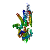
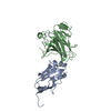
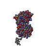
- PDB-3u43: Crystal structure of the colicin E2 DNase-Im2 complex -
+
Open data
ID or keywords:
Loading...
-
Basic information
Entry
Database: PDB / ID: 3u43
Title
Crystal structure of the colicin E2 DNase-Im2 complex
Components
Colicin-E2
Colicin-E2 immunity protein
Keywords
PROTEIN BINDING / protein-protein complex / DNase / high affinity
Function / homology
Function and homology information
extrachromosomal circular DNA / bacteriocin immunity / toxic substance binding / endonuclease activity / killing of cells of another organism / Hydrolases; Acting on ester bonds / defense response to bacterium / metal ion binding Similarity search - Function
Colicin E7 immunity protein; Chain B, fragment: Endonuclease domain / Colicin/pyocin, DNase domain / Colicin E immunity protein / Colicin immunity protein/pyocin immunity protein / Colicin E immunity protein superfamily / Colicin immunity protein / pyocin immunity protein / Colicin/Pyocin-S2, DNase domain / Colicin/pyocin, DNase domain superfamily / Colicin, receptor domain / Coiled-coil receptor-binding R-domain of colicin E2 ...Colicin E7 immunity protein; Chain B, fragment: Endonuclease domain / Colicin/pyocin, DNase domain / Colicin E immunity protein / Colicin immunity protein/pyocin immunity protein / Colicin E immunity protein superfamily / Colicin immunity protein / pyocin immunity protein / Colicin/Pyocin-S2, DNase domain / Colicin/pyocin, DNase domain superfamily / Colicin, receptor domain / Coiled-coil receptor-binding R-domain of colicin E2 / Cloacin colicin family / Colicin-like bacteriocin tRNase domain / Pyosin/cloacin translocation domain / Pyosin/cloacin translocation domain superfamily / HNH nucleases / His-Me finger superfamily / HNH nuclease / Non-ribosomal Peptide Synthetase Peptidyl Carrier Protein; Chain A / Alpha-Beta Complex / Orthogonal Bundle / Mainly Alpha / Alpha Beta Similarity search - Domain/homology
Monochromator: Si(111) / Protocol: SINGLE WAVELENGTH / Monochromatic (M) / Laue (L): M / Scattering type: x-ray
Radiation wavelength
Wavelength: 0.8726 Å / Relative weight: 1
Reflection
Resolution: 1.714→121.814 Å / Num. obs: 23549 / % possible obs: 99.1 % / Redundancy: 6.9 % / Rsym value: 0.084 / Net I/σ(I): 14.4
Reflection shell
Diffraction-ID: 1
Resolution (Å)
Redundancy (%)
Rmerge(I) obs
Mean I/σ(I) obs
Num. measured all
Num. unique all
Rsym value
% possible all
1.71-1.81
5.2
0.427
1.8
16598
3174
0.427
93.9
1.81-1.92
7.3
0.287
2.7
23434
3226
0.287
100
1.92-2.05
7.3
0.184
4.2
22213
3057
0.184
100
2.05-2.21
7.3
0.13
5.8
20609
2832
0.13
100
2.21-2.42
7.3
0.101
7.3
19033
2625
0.101
100
2.42-2.71
7.2
0.082
8.8
17267
2383
0.082
100
2.71-3.13
7.2
0.071
9.6
15222
2119
0.071
100
3.13-3.83
7.1
0.059
11.1
12945
1820
0.059
100
3.83-5.42
6.9
0.049
12.6
10011
1445
0.049
100
5.42-48.813
6.3
0.04
15.5
5476
868
0.04
99.9
-
Phasing
Phasing
Method: molecular replacement
Phasing MR
Highest resolution
Lowest resolution
Rotation
1.95 Å
48.81 Å
Translation
1.95 Å
48.81 Å
-
Processing
Software
Name
Version
Classification
NB
SCALA
3.3.15
datascaling
MOLREP
phasing
REFMAC
refinement
PDB_EXTRACT
3.1
dataextraction
MxCuBE
datacollection
XDS
datareduction
Refinement
Method to determine structure: MOLECULAR REPLACEMENT / Resolution: 1.72→48.81 Å / Cor.coef. Fo:Fc: 0.963 / Cor.coef. Fo:Fc free: 0.946 / WRfactor Rfree: 0.1939 / WRfactor Rwork: 0.1526 / Occupancy max: 1 / Occupancy min: 0.4 / FOM work R set: 0.8812 / SU B: 2.145 / SU ML: 0.071 / SU R Cruickshank DPI: 0.115 / SU Rfree: 0.1118 / Cross valid method: THROUGHOUT / σ(F): 0 / ESU R Free: 0.112 / Stereochemistry target values: MAXIMUM LIKELIHOOD Details: HYDROGENS HAVE BEEN USED IF PRESENT IN THE INPUT. U VALUES: REFINED INDIVIDUALLY
Rfactor
Num. reflection
% reflection
Selection details
Rfree
0.2025
1206
5.1 %
RANDOM
Rwork
0.1616
-
-
-
all
0.1638
23419
-
-
obs
0.1638
23419
99.71 %
-
Solvent computation
Ion probe radii: 0.8 Å / Shrinkage radii: 0.8 Å / VDW probe radii: 1.2 Å / Solvent model: MASK
In the structure databanks used in Yorodumi, some data are registered as the other names, "COVID-19 virus" and "2019-nCoV". Here are the details of the virus and the list of structure data.
Jan 31, 2019. EMDB accession codes are about to change! (news from PDBe EMDB page)
EMDB accession codes are about to change! (news from PDBe EMDB page)
The allocation of 4 digits for EMDB accession codes will soon come to an end. Whilst these codes will remain in use, new EMDB accession codes will include an additional digit and will expand incrementally as the available range of codes is exhausted. The current 4-digit format prefixed with “EMD-” (i.e. EMD-XXXX) will advance to a 5-digit format (i.e. EMD-XXXXX), and so on. It is currently estimated that the 4-digit codes will be depleted around Spring 2019, at which point the 5-digit format will come into force.
The EM Navigator/Yorodumi systems omit the EMD- prefix.
Related info.:Q: What is EMD? / ID/Accession-code notation in Yorodumi/EM Navigator
Yorodumi is a browser for structure data from EMDB, PDB, SASBDB, etc.
This page is also the successor to EM Navigator detail page, and also detail information page/front-end page for Omokage search.
The word "yorodu" (or yorozu) is an old Japanese word meaning "ten thousand". "mi" (miru) is to see.
Related info.:EMDB / PDB / SASBDB / Comparison of 3 databanks / Yorodumi Search / Aug 31, 2016. New EM Navigator & Yorodumi / Yorodumi Papers / Jmol/JSmol / Function and homology information / Changes in new EM Navigator and Yorodumi
 Movie
Movie Controller


 Open data
 Basic information
 Components
 Keywords
 PROTEIN BINDING / protein-protein complex /
  Function and homology information
 Authors
 Citation
 Structure visualization
 Downloads & links
 Other downloads


 PDBj
PDBj
 Assembly


 Mass: 65.409 Da / Num. of mol.: 1 / Source method: obtained synthetically / Formula: Zn
 Mass: 40.078 Da / Num. of mol.: 1 / Source method: obtained synthetically / Formula: Ca
 Mass: 18.015 Da / Num. of mol.: 319 / Source method: isolated from a natural source / Formula: H2O
 Sample preparation
 / Beamline: ID23-2 / Wavelength: 0.8726 Å
 Processing