[English] 日本語
Yorodumi
- PDB-3u33: Crystal Structure of the E. coli adaptive response protein AidB i... -

+
Open data


ID or keywords:

Loading...

-
Basic information

Entry
Database: PDB / ID: 3u33
TitleCrystal Structure of the E. coli adaptive response protein AidB in the space group P3(2)
ComponentsPutative acyl-CoA dehydrogenase AidB
KeywordsOXIDOREDUCTASE / acyl-coenzyme A dehydrogenase / protective function in the presence of alkylating agents / DNA binding / FAD binding
Function / homology
Function and homology information


isovaleryl-CoA dehydrogenase activity / Oxidoreductases; Acting on the CH-CH group of donors; With unknown physiological acceptors / acyl-CoA dehydrogenase activity / sequence-specific DNA binding / negative regulation of DNA-templated transcription / DNA damage response / DNA binding / identical protein binding / cytoplasm
Similarity search - Function
Butyryl-CoA Dehydrogenase, subunit A; domain 2 - #20 / Single alpha-helices involved in coiled-coils or other helix-helix interfaces - #600 / Putative acyl-CoA dehydrogenase AidB / Adaptive response protein AidB, N-terminal / Adaptive response protein AidB N-terminal domain / Acyl-CoA dehydrogenases signature 1. / Acyl-CoA dehydrogenases signature 2. / Acyl-CoA dehydrogenase, conserved site / Butyryl-CoA Dehydrogenase, subunit A; domain 2 / Acyl-CoA dehydrogenase/oxidase C-terminal ...Butyryl-CoA Dehydrogenase, subunit A; domain 2 - #20 / Single alpha-helices involved in coiled-coils or other helix-helix interfaces - #600 / Putative acyl-CoA dehydrogenase AidB / Adaptive response protein AidB, N-terminal / Adaptive response protein AidB N-terminal domain / Acyl-CoA dehydrogenases signature 1. / Acyl-CoA dehydrogenases signature 2. / Acyl-CoA dehydrogenase, conserved site / Butyryl-CoA Dehydrogenase, subunit A; domain 2 / Acyl-CoA dehydrogenase/oxidase C-terminal / Acyl-CoA dehydrogenase, C-terminal domain / Acyl-CoA oxidase/dehydrogenase, middle domain / Acyl-CoA dehydrogenase, middle domain / Acyl-CoA dehydrogenase/oxidase, N-terminal and middle domain superfamily / Acyl-CoA dehydrogenase-like, C-terminal / Single alpha-helices involved in coiled-coils or other helix-helix interfaces / Helix non-globular / Special / Beta Barrel / Mainly Beta
Similarity search - Domain/homology
FLAVIN-ADENINE DINUCLEOTIDE / Putative acyl-CoA dehydrogenase AidB
Similarity search - Component
Biological speciesEscherichia coli K-12 (bacteria)
MethodX-RAY DIFFRACTION / SYNCHROTRON / MOLECULAR REPLACEMENT / Resolution: 2.8 Å
AuthorsWong, C. / Jost, M. / Drennan, C.L.
CitationJournal: Biochemistry / Year: 2011
Title: Flavin-induced oligomerization in Escherichia coli adaptive response protein AidB.
Authors: Hamill, M.J. / Jost, M. / Wong, C. / Elliott, S.J. / Drennan, C.L.
History
DepositionOct 4, 2011Deposition site: RCSB / Processing site: RCSB
Revision 1.0Nov 2, 2011Provider: repository / Type: Initial release
Revision 1.1Jan 9, 2013Group: Database references
Revision 1.2Oct 25, 2017Group: Author supporting evidence / Category: pdbx_struct_assembly_auth_evidence
Revision 1.3Sep 13, 2023Group: Data collection / Database references ...Data collection / Database references / Derived calculations / Refinement description
Category: chem_comp_atom / chem_comp_bond ...chem_comp_atom / chem_comp_bond / database_2 / pdbx_initial_refinement_model / struct_site
Item: _database_2.pdbx_DOI / _database_2.pdbx_database_accession ..._database_2.pdbx_DOI / _database_2.pdbx_database_accession / _struct_site.pdbx_auth_asym_id / _struct_site.pdbx_auth_comp_id / _struct_site.pdbx_auth_seq_id

-
Structure visualization

Structure viewerMolecule:
MolmilJmol/JSmol

Downloads & links

-
Assembly

Deposited unit
A: Putative acyl-CoA dehydrogenase AidB
B: Putative acyl-CoA dehydrogenase AidB
C: Putative acyl-CoA dehydrogenase AidB
D: Putative acyl-CoA dehydrogenase AidB
E: Putative acyl-CoA dehydrogenase AidB
F: Putative acyl-CoA dehydrogenase AidB
G: Putative acyl-CoA dehydrogenase AidB
H: Putative acyl-CoA dehydrogenase AidB
I: Putative acyl-CoA dehydrogenase AidB
J: Putative acyl-CoA dehydrogenase AidB
K: Putative acyl-CoA dehydrogenase AidB
L: Putative acyl-CoA dehydrogenase AidB
hetero molecules


Theoretical massNumber of molelcules
Total (without water)737,80636
Polymers727,95412
Non-polymers9,85224
Water5,350297
1
A: Putative acyl-CoA dehydrogenase AidB
B: Putative acyl-CoA dehydrogenase AidB
C: Putative acyl-CoA dehydrogenase AidB
D: Putative acyl-CoA dehydrogenase AidB
hetero molecules


Theoretical massNumber of molelcules
Total (without water)245,93512
Polymers242,6514
Non-polymers3,2848
Water724
TypeNameSymmetry operationNumber
identity operation1_555x,y,z1
Buried area26740 Å2
ΔGint-152 kcal/mol
Surface area67980 Å2
MethodPISA
2
E: Putative acyl-CoA dehydrogenase AidB
F: Putative acyl-CoA dehydrogenase AidB
G: Putative acyl-CoA dehydrogenase AidB
H: Putative acyl-CoA dehydrogenase AidB
hetero molecules


Theoretical massNumber of molelcules
Total (without water)245,93512
Polymers242,6514
Non-polymers3,2848
Water724
TypeNameSymmetry operationNumber
identity operation1_555x,y,z1
Buried area26880 Å2
ΔGint-154 kcal/mol
Surface area67900 Å2
MethodPISA
3
I: Putative acyl-CoA dehydrogenase AidB
J: Putative acyl-CoA dehydrogenase AidB
K: Putative acyl-CoA dehydrogenase AidB
L: Putative acyl-CoA dehydrogenase AidB
hetero molecules


Theoretical massNumber of molelcules
Total (without water)245,93512
Polymers242,6514
Non-polymers3,2848
Water724
TypeNameSymmetry operationNumber
identity operation1_555x,y,z1
Buried area26740 Å2
ΔGint-157 kcal/mol
Surface area67960 Å2
MethodPISA
Unit cell
Length a, b, c (Å)179.720, 179.720, 204.237
Angle α, β, γ (deg.)90.00, 90.00, 120.00
Int Tables number145
Space group name H-MP32
Noncrystallographic symmetry (NCS)NCS domain:
IDEns-ID
11
21
31
41
51
61

NCS domain segments:
Dom-IDComponent-IDEns-IDSelection details
111(chain A or chain B) and (resseq 2:162 or resseq...
211(chain D or chain C) and (resseq 2:162 or resseq...
311(chain E or chain F) and (resseq 2:162 or resseq...
411(chain H or chain G) and (resseq 2:162 or resseq...
511(chain I or chain J) and (resseq 2:162 or resseq...
611(chain L or chain K) and (resseq 2:162 or resseq...
Details
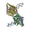
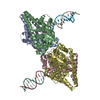
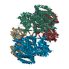
Three tetramers in the asymmetric unit, composed of A+B+C+D, E+F+G+H, and I+J+K+L. Tetrameric assembly independently confirmed by analytical ultracentrifugation.

-
Components

#1: Protein
Putative acyl-CoA dehydrogenase AidB / adaptive response protein AidB


Mass: 60662.824 Da / Num. of mol.: 12
Source method: isolated from a genetically manipulated source
Source: (gene. exp.) Escherichia coli K-12 (bacteria) / Strain: AB1157 / Gene: aidB, b4187, JW5867 / Plasmid: pET28a / Production host: Escherichia coli (E. coli) / Strain (production host): BL21(DE3)
References: UniProt: P33224, Oxidoreductases; Acting on the CH-CH group of donors; With unknown physiological acceptors
#2: Chemical
ChemComp-FAD / FLAVIN-ADENINE DINUCLEOTIDE / Flavin adenine dinucleotide


Mass: 785.550 Da / Num. of mol.: 12 / Source method: obtained synthetically / Formula: C27H33N9O15P2 / Comment: FAD*YM
#3: Chemical
ChemComp-CL / CHLORIDE ION / Chloride


Mass: 35.453 Da / Num. of mol.: 12 / Source method: obtained synthetically / Formula: Cl
#4: Water ChemComp-HOH / water / Water


Mass: 18.015 Da / Num. of mol.: 297 / Source method: isolated from a natural source / Formula: H2O

-
Experimental details

-
Experiment

ExperimentMethod: X-RAY DIFFRACTION / Number of used crystals: 1

-
Sample preparation

CrystalDensity Matthews: 2.62 Å3/Da / Density % sol: 52.98 %
Crystal growTemperature: 298 K / Method: vapor diffusion, hanging drop / pH: 7.5
Details: 20% (v/v) ethanol, 0.1 M HEPES pH 7.5, 0.2 M magnesium chloride, VAPOR DIFFUSION, HANGING DROP, temperature 298K

-
Data collection

DiffractionMean temperature: 100 K
Diffraction sourceSource: SYNCHROTRON / Site: APS / Beamline: 24-ID-C / Wavelength: 1 Å
DetectorType: ADSC QUANTUM 315 / Detector: CCD / Date: Mar 5, 2008
RadiationMonochromator: Cryogenically-cooled single crystal Si(220) side bounce monochromator
Protocol: SINGLE WAVELENGTH / Monochromatic (M) / Laue (L): M / Scattering type: x-ray
Radiation wavelengthWavelength: 1 Å / Relative weight: 1
ReflectionResolution: 2.8→50 Å / Num. all: 174735 / Num. obs: 174735 / % possible obs: 96.2 % / Observed criterion σ(I): -3 / Redundancy: 6.1 % / Biso Wilson estimate: 45.1 Å2 / Rsym value: 0.095 / Net I/σ(I): 14.1
Reflection shellResolution: 2.8→2.9 Å / Redundancy: 5.1 % / Mean I/σ(I) obs: 3.33 / Num. unique all: 17687 / Rsym value: 0.457 / % possible all: 97.4

-
Processing

Software
NameVersionClassification
ADSCQuantumdata collection
PHASERphasing
PHENIX(phenix.refine: 1.7.1_743)refinement
HKL-2000data reduction
HKL-2000data scaling
RefinementMethod to determine structure: MOLECULAR REPLACEMENT
Starting model: PDB ENTRY 3DJL
Resolution: 2.8→46.254 Å / SU ML: 0.78 / Cross valid method: THROUGHOUT / σ(F): 1 / Phase error: 23.85 / Stereochemistry target values: ML
RfactorNum. reflection% reflectionSelection details
Rfree0.2289 8633 5.01 %random
Rwork0.2045 ---
obs0.2058 172442 94.99 %-
all-174695 --
Solvent computationShrinkage radii: 0.9 Å / VDW probe radii: 1.11 Å / Solvent model: FLAT BULK SOLVENT MODEL / Bsol: 8.302 Å2 / ksol: 0.278 e/Å3
Displacement parameters
Baniso -1Baniso -2Baniso -3
1-1.2626 Å2-0 Å2-0 Å2
2--1.2626 Å2-0 Å2
3----0.8276 Å2
Refinement stepCycle: LAST / Resolution: 2.8→46.254 Å
ProteinNucleic acidLigandSolventTotal
Num. atoms49649 0 648 297 50594
Refine LS restraints
Refine-IDTypeDev idealNumber
X-RAY DIFFRACTIONf_bond_d0.00551457
X-RAY DIFFRACTIONf_angle_d0.82869861
X-RAY DIFFRACTIONf_dihedral_angle_d12.75218559
X-RAY DIFFRACTIONf_chiral_restr0.0547635
X-RAY DIFFRACTIONf_plane_restr0.0039037
Refine LS restraints NCS
Ens-IDDom-IDAuth asym-IDNumberRefine-IDTypeRms dev position (Å)
11A7875X-RAY DIFFRACTIONPOSITIONAL
12D7875X-RAY DIFFRACTIONPOSITIONAL0.051
13E7884X-RAY DIFFRACTIONPOSITIONAL0.044
14H7888X-RAY DIFFRACTIONPOSITIONAL0.047
15I7904X-RAY DIFFRACTIONPOSITIONAL0.047
16L7849X-RAY DIFFRACTIONPOSITIONAL0.058
LS refinement shell
Resolution (Å)Rfactor RfreeNum. reflection RfreeRfactor RworkNum. reflection RworkRefine-ID% reflection obs (%)
2.8004-2.83220.35172720.30915385X-RAY DIFFRACTION94
2.8322-2.86550.32873000.29655456X-RAY DIFFRACTION95
2.8655-2.90050.32773220.29365443X-RAY DIFFRACTION95
2.9005-2.93720.32012890.28645440X-RAY DIFFRACTION96
2.9372-2.97580.29642860.27475553X-RAY DIFFRACTION96
2.9758-3.01660.28342860.25875550X-RAY DIFFRACTION97
3.0166-3.05970.31052710.26055674X-RAY DIFFRACTION97
3.0597-3.10530.282920.25065586X-RAY DIFFRACTION98
3.1053-3.15380.26993130.24175654X-RAY DIFFRACTION98
3.1538-3.20550.2742980.23745638X-RAY DIFFRACTION98
3.2055-3.26080.24552680.2235620X-RAY DIFFRACTION98
3.2608-3.32010.23663080.21925677X-RAY DIFFRACTION99
3.3201-3.38390.25093120.20865646X-RAY DIFFRACTION99
3.3839-3.4530.24043060.22255681X-RAY DIFFRACTION98
3.453-3.5280.22742810.20665632X-RAY DIFFRACTION98
3.528-3.61010.22132830.21145672X-RAY DIFFRACTION98
3.6101-3.70030.5042680.49711564X-RAY DIFFRACTION35
3.7003-3.80030.2232530.20044970X-RAY DIFFRACTION88
3.8003-3.91210.23872740.20325378X-RAY DIFFRACTION94
3.9121-4.03830.21112880.16535323X-RAY DIFFRACTION93
4.0383-4.18250.1683310.15265786X-RAY DIFFRACTION100
4.1825-4.34990.16452970.14695700X-RAY DIFFRACTION100
4.3499-4.54770.19532990.14615729X-RAY DIFFRACTION100
4.5477-4.78720.18172950.15435739X-RAY DIFFRACTION100
4.7872-5.08680.19923170.16845713X-RAY DIFFRACTION100
5.0868-5.4790.19372980.18455732X-RAY DIFFRACTION100
5.479-6.02930.21693230.19015750X-RAY DIFFRACTION100
6.0293-6.89920.23443070.19885763X-RAY DIFFRACTION100
6.8992-8.68290.1742910.15895705X-RAY DIFFRACTION100
8.6829-46.260.19453050.18195650X-RAY DIFFRACTION98
Refinement TLS params.

Method: refined / Refine-ID: X-RAY DIFFRACTION

IDL112)L122)L132)L222)L232)L332)S11 (Å °)S12 (Å °)S13 (Å °)S21 (Å °)S22 (Å °)S23 (Å °)S31 (Å °)S32 (Å °)S33 (Å °)T112)T122)T132)T222)T232)T332)Origin x (Å)Origin y (Å)Origin z (Å)
10.3718-0.1588-0.01770.4984-0.00470.56610.0222-0.1122-0.21250.18480.05430.13420.15610.12010.0165-0.2294-0.0797-0.03350.02970.08520.0371-5.78314.329-67.0106
20.67710.0384-0.05920.4085-0.08180.5906-0.06740.0564-0.10120.11090.0980.0592-0.0029-0.09090.00190.02980.08470.01780.1211-0.08670.037479.994438.9685-74.3771
30.3805-0.08610.23460.5358-0.12260.86450.47960.16490.2903-0.0106-0.04560.0151-0.0371-0.10690.0885-0.4707-0.35620.3063-0.44080.2166-0.239615.0837101.4308-72.1579
Refinement TLS group
IDRefine-IDRefine TLS-IDSelection details
1X-RAY DIFFRACTION1((chain A or chain B or chain C or chain D)
2X-RAY DIFFRACTION2((chain E or chain F or chain G or chain H)
3X-RAY DIFFRACTION3((chain I or chain J or chain K or chain L)

+
About Yorodumi

-
News

-
Feb 9, 2022. New format data for meta-information of EMDB entries

New format data for meta-information of EMDB entries

  • Version 3 of the EMDB header file is now the official format.
  • The previous official version 1.9 will be removed from the archive.

Related info.:EMDB header

External links:wwPDB to switch to version 3 of the EMDB data model

-
Aug 12, 2020. Covid-19 info

Covid-19 info

URL: https://pdbj.org/emnavi/covid19.php

New page: Covid-19 featured information page in EM Navigator.

Related info.:Covid-19 info / Mar 5, 2020. Novel coronavirus structure data

+
Mar 5, 2020. Novel coronavirus structure data

Novel coronavirus structure data

Related info.:Yorodumi Speices / Aug 12, 2020. Covid-19 info

External links:COVID-19 featured content - PDBj / Molecule of the Month (242):Coronavirus Proteases

+
Jan 31, 2019. EMDB accession codes are about to change! (news from PDBe EMDB page)

EMDB accession codes are about to change! (news from PDBe EMDB page)

  • The allocation of 4 digits for EMDB accession codes will soon come to an end. Whilst these codes will remain in use, new EMDB accession codes will include an additional digit and will expand incrementally as the available range of codes is exhausted. The current 4-digit format prefixed with “EMD-” (i.e. EMD-XXXX) will advance to a 5-digit format (i.e. EMD-XXXXX), and so on. It is currently estimated that the 4-digit codes will be depleted around Spring 2019, at which point the 5-digit format will come into force.
  • The EM Navigator/Yorodumi systems omit the EMD- prefix.

Related info.:Q: What is EMD? / ID/Accession-code notation in Yorodumi/EM Navigator

External links:EMDB Accession Codes are Changing Soon! / Contact to PDBj

+
Jul 12, 2017. Major update of PDB

Major update of PDB

  • wwPDB released updated PDB data conforming to the new PDBx/mmCIF dictionary.
  • This is a major update changing the version number from 4 to 5, and with Remediation, in which all the entries are updated.
  • In this update, many items about electron microscopy experimental information are reorganized (e.g. em_software).
  • Now, EM Navigator and Yorodumi are based on the updated data.

External links:wwPDB Remediation / Enriched Model Files Conforming to OneDep Data Standards Now Available in the PDB FTP Archive

-
Yorodumi

Thousand views of thousand structures

  • Yorodumi is a browser for structure data from EMDB, PDB, SASBDB, etc.
  • This page is also the successor to EM Navigator detail page, and also detail information page/front-end page for Omokage search.
  • The word "yorodu" (or yorozu) is an old Japanese word meaning "ten thousand". "mi" (miru) is to see.

Related info.:EMDB / PDB / SASBDB / Comparison of 3 databanks / Yorodumi Search / Aug 31, 2016. New EM Navigator & Yorodumi / Yorodumi Papers / Jmol/JSmol / Function and homology information / Changes in new EM Navigator and Yorodumi

Read more