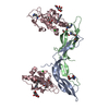
Entry Database : PDB / ID : 3u2pTitle Crystal structure of N-terminal three extracellular domains of ErbB4/Her4 Receptor tyrosine-protein kinase erbB-4 Keywords / / / Function / homology Function Domain/homology Component
/ / / / / / / / / / / / / / / / / / / / / / / / / / / / / / / / / / / / / / / / / / / / / / / / / / / / / / / / / / / / / / / / / / / / / / / / / / / / / / / / / / / / / / / / / / / / / / / / / / / / / / / / / / / / / / / / / / / / / / / / / / / / / / / / / / / / Biological species Homo sapiens (human)Method / / Resolution : 2.57 Å Authors Liu, P. / Bouyain, S. / Elgenbrot, C. / Leahy, D.J. Journal : Protein Sci. / Year : 2012Title : The ErbB4 extracellular region retains a tethered-like conformation in the absence of the tether.Authors : Liu, P. / Bouyain, S. / Eigenbrot, C. / Leahy, D.J. History Deposition Oct 4, 2011 Deposition site / Processing site Revision 1.0 Nov 9, 2011 Provider / Type Revision 1.1 Oct 3, 2012 Group Revision 1.2 Jul 29, 2020 Group / Derived calculations / Structure summaryCategory chem_comp / entity ... chem_comp / entity / pdbx_chem_comp_identifier / pdbx_entity_nonpoly / struct_conn / struct_site / struct_site_gen Item _chem_comp.name / _chem_comp.type ... _chem_comp.name / _chem_comp.type / _entity.pdbx_description / _pdbx_entity_nonpoly.name / _struct_conn.pdbx_dist_value / _struct_conn.pdbx_leaving_atom_flag / _struct_conn.pdbx_role / _struct_conn.ptnr1_auth_seq_id / _struct_conn.ptnr1_label_seq_id / _struct_conn.ptnr2_auth_seq_id / _struct_conn.ptnr2_label_asym_id Description / Provider / Type Revision 1.3 Sep 13, 2023 Group Data collection / Database references ... Data collection / Database references / Refinement description / Structure summary Category chem_comp / chem_comp_atom ... chem_comp / chem_comp_atom / chem_comp_bond / database_2 / pdbx_initial_refinement_model Item / _database_2.pdbx_DOI / _database_2.pdbx_database_accession
Show all Show less
 Movie
Movie Controller

 Yorodumi
 Open data
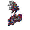
 Basic information
 Components
 Keywords
 Transferase /
  Function and homology information
 Authors
 Citation
 Structure visualization
 Downloads & links
 Other downloads
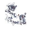


 PDBj
PDBj


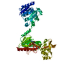
 Assembly


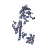
 Type: D-saccharide, beta linking / Mass: 221.208 Da / Num. of mol.: 5
 Mass: 18.015 Da / Num. of mol.: 86 / Source method: isolated from a natural source / Formula: H2O
 Sample preparation
 Processing