 Movie
Movie Controller

[English] 日本語
 Yorodumi
- PDB-3u2g: Crystal structure of the C-terminal DUF1608 domain of the Methano... -
+ Open data

- Basic information
| Entry | Database: PDB / ID: 3u2g | ||||||
|---|---|---|---|---|---|---|---|
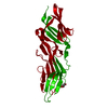
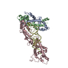
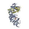
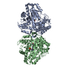
| Title | Crystal structure of the C-terminal DUF1608 domain of the Methanosarcina acetivorans S-layer (MA0829) protein | ||||||
 Components Components | S-layer protein MA0829 | ||||||
 Keywords Keywords | STRUCTURAL PROTEIN / DUF1608 domain / cell envelop / surface layer / S-layer / UNKNOWN FUNCTION | ||||||
| Function / homology |  Function and homology informationS-layer / cell wall organization / extracellular region / identical protein binding / plasma membrane Function and homology informationS-layer / cell wall organization / extracellular region / identical protein binding / plasma membraneSimilarity search - Function | ||||||
| Biological species |  Methanosarcina acetivorans (archaea) Methanosarcina acetivorans (archaea) | ||||||
| Method | X-RAY DIFFRACTION / SYNCHROTRON / SAD / Resolution: 2.3 Å | ||||||
 Authors Authors | Chan, S. / Phan, T. / Ahn, C.J. / Shin, A. / Rohlin, L. / Gunsalus, R.P. / Arbing, M.A. | ||||||
 Citation Citation | Journal: Proc.Natl.Acad.Sci.USA / Year: 2012 Title: Structure of the surface layer of the methanogenic archaean Methanosarcina acetivorans. Authors: Arbing, M.A. / Chan, S. / Shin, A. / Phan, T. / Ahn, C.J. / Rohlin, L. / Gunsalus, R.P. | ||||||
| History |
|
- Structure visualization
| Structure viewer | Molecule: MolmilJmol/JSmol |
|---|
- Downloads & links
-Download
| PDBx/mmCIF format | 3u2g.cif.gz | 118.7 KB | Display | PDBx/mmCIF format |
|---|---|---|---|---|
| PDB format | pdb3u2g.ent.gz | 98.3 KB | Display | PDB format |
| PDBx/mmJSON format | 3u2g.json.gz | Tree view | PDBx/mmJSON format | |
| Others |  Other downloads Other downloads |
-Validation report
| Arichive directory | https://data.pdbj.org/pub/pdb/validation_reports/u2/3u2gftp://data.pdbj.org/pub/pdb/validation_reports/u2/3u2g | HTTPS FTP |
|---|
-Related structure data


-Links
 PDBj
PDBj
- Assembly
| Deposited unit | 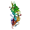
| ||||||||
|---|---|---|---|---|---|---|---|---|---|
| 1 | 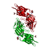
| ||||||||
| Unit cell |
| ||||||||
| Details | the full-length natural protein consists of a tandem repeats of two DUF1608 domains of highly similar sequences |
-Components
| #1: Protein | Mass: 32389.953 Da / Num. of mol.: 1 / Fragment: C-terminal DUF1608 domain Source method: isolated from a genetically manipulated source Source: (gene. exp.) Methanosarcina acetivorans (archaea) / Strain: C2A / Gene: MA0829, MA_0829 / Plasmid: pET22b / Production host:  Escherichia coli (E. coli) / Strain (production host): BL21 Rosetta (DE3) / References: UniProt: Q8TSG7 Escherichia coli (E. coli) / Strain (production host): BL21 Rosetta (DE3) / References: UniProt: Q8TSG7 | ||
|---|---|---|---|
| #2: Chemical | ChemComp-CIT / Citric acid  Mass: 192.124 Da / Num. of mol.: 1 / Source method: obtained synthetically / Formula: C6H8O7 Mass: 192.124 Da / Num. of mol.: 1 / Source method: obtained synthetically / Formula: C6H8O7 | ||
| #3: Chemical | ChemComp-NH4 / Ammonium  Mass: 18.038 Da / Num. of mol.: 1 / Source method: obtained synthetically / Formula: H4N Mass: 18.038 Da / Num. of mol.: 1 / Source method: obtained synthetically / Formula: H4N | ||
| #4: Chemical | Glycerol  Mass: 92.094 Da / Num. of mol.: 2 / Source method: obtained synthetically / Formula: C3H8O3 Mass: 92.094 Da / Num. of mol.: 2 / Source method: obtained synthetically / Formula: C3H8O3#5: Water | ChemComp-HOH / | Water Mass: 18.015 Da / Num. of mol.: 168 / Source method: isolated from a natural source / Formula: H2O Mass: 18.015 Da / Num. of mol.: 168 / Source method: isolated from a natural source / Formula: H2O |
-Experimental details
-Experiment
| Experiment | Method: X-RAY DIFFRACTION / Number of used crystals: 1 |
|---|
- Sample preparation
| Crystal | Density Matthews: 2.9 Å3/Da / Density % sol: 57.62 % |
|---|---|
| Crystal grow | Temperature: 293 K / Method: vapor diffusion, hanging drop / pH: 7.4 Details: 0.4 M ammonium citrate, 20% PEG3350, pH 7.4, VAPOR DIFFUSION, HANGING DROP, temperature 293K |
-Data collection
| Diffraction | Mean temperature: 100 K |
|---|---|
| Diffraction source | Source: SYNCHROTRON / Site: APS  / Beamline: 24-ID-C / Wavelength: 0.979028 Å / Beamline: 24-ID-C / Wavelength: 0.979028 Å |
| Detector | Type: ADSC QUANTUM 315 / Detector: CCD / Date: Feb 20, 2010 |
| Radiation | Monochromator: Si(111) double crystal monochromator (Kohzu HLD8-24 Monochromator) Protocol: SINGLE WAVELENGTH / Monochromatic (M) / Laue (L): M / Scattering type: x-ray |
| Radiation wavelength | Wavelength: 0.979028 Å / Relative weight: 1 |
| Reflection | Resolution: 2.3→90 Å / Num. obs: 17588 / % possible obs: 99.9 % / Redundancy: 10.4 % / Rsym value: 0.092 / Net I/σ(I): 21.1 |
| Reflection shell | Resolution: 2.3→2.38 Å / Redundancy: 10.7 % / Mean I/σ(I) obs: 6.2 / Num. unique all: 1715 / Rsym value: 0.494 / % possible all: 100 |
- Processing
| Software |
| |||||||||||||||||||||||||||||||||||||||||||||||||||||||||||||||||||||||||||||||||||||
|---|---|---|---|---|---|---|---|---|---|---|---|---|---|---|---|---|---|---|---|---|---|---|---|---|---|---|---|---|---|---|---|---|---|---|---|---|---|---|---|---|---|---|---|---|---|---|---|---|---|---|---|---|---|---|---|---|---|---|---|---|---|---|---|---|---|---|---|---|---|---|---|---|---|---|---|---|---|---|---|---|---|---|---|---|---|---|
| Refinement | Method to determine structure: SAD / Resolution: 2.3→90 Å / Cor.coef. Fo:Fc: 0.944 / Cor.coef. Fo:Fc free: 0.924 / Occupancy max: 1 / Occupancy min: 0.5 / SU B: 10.602 / SU ML: 0.118 / Cross valid method: THROUGHOUT / σ(F): 0 / ESU R Free: 0.195 / Stereochemistry target values: MAXIMUM LIKELIHOOD Details: HYDROGENS HAVE BEEN ADDED IN THE RIDING POSITIONS U VALUES : RESIDUAL ONLY
| |||||||||||||||||||||||||||||||||||||||||||||||||||||||||||||||||||||||||||||||||||||
| Solvent computation | Ion probe radii: 0.8 Å / Shrinkage radii: 0.8 Å / VDW probe radii: 1.4 Å / Solvent model: MASK | |||||||||||||||||||||||||||||||||||||||||||||||||||||||||||||||||||||||||||||||||||||
| Displacement parameters | Biso max: 69.78 Å2 / Biso mean: 27.182 Å2 / Biso min: 8.81 Å2
| |||||||||||||||||||||||||||||||||||||||||||||||||||||||||||||||||||||||||||||||||||||
| Refinement step | Cycle: LAST / Resolution: 2.3→90 Å
| |||||||||||||||||||||||||||||||||||||||||||||||||||||||||||||||||||||||||||||||||||||
| Refine LS restraints |
| |||||||||||||||||||||||||||||||||||||||||||||||||||||||||||||||||||||||||||||||||||||
| LS refinement shell | Resolution: 2.3→2.36 Å / Total num. of bins used: 20
| |||||||||||||||||||||||||||||||||||||||||||||||||||||||||||||||||||||||||||||||||||||
| Refinement TLS params. | Method: refined / Origin x: 12.7909 Å / Origin y: 45.2377 Å / Origin z: 19.8265 Å
|
