 Movie
Movie Controller

[English] 日本語
 Yorodumi
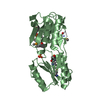
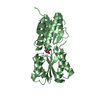
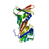
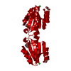
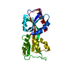
- PDB-3u21: Crystal structure of a Fragment of Nuclear factor related to kapp... -
+ Open data

- Basic information
| Entry | Database: PDB / ID: 3u21 | ||||||
|---|---|---|---|---|---|---|---|
| Title | Crystal structure of a Fragment of Nuclear factor related to kappa-B-binding protein (residues 370-495) (NFRKB) from Homo sapiens at 2.18 A resolution | ||||||
 Components Components | Nuclear factor related to kappa-B-binding protein | ||||||
 Keywords Keywords |  Transcription regulation / DNA BINDING / DNA/RNA-binding 3-helical bundle / winged-HTH domain / Structural Genomics / Joint Center for Structural Genomics / JCSG / Protein Structure Initiative / PSI-BIOLOGY / DNA BINDING PROTEIN / Partnership for Stem Cell Biology / STEMCELL Transcription regulation / DNA BINDING / DNA/RNA-binding 3-helical bundle / winged-HTH domain / Structural Genomics / Joint Center for Structural Genomics / JCSG / Protein Structure Initiative / PSI-BIOLOGY / DNA BINDING PROTEIN / Partnership for Stem Cell Biology / STEMCELL | ||||||
| Function / homology |  Function and homology information Function and homology informationregulation of DNA strand elongation / positive regulation of telomere maintenance in response to DNA damage / Ino80 complex / regulation of chromosome organization / regulation of DNA replication / regulation of embryonic development / regulation of DNA repair / positive regulation of DNA repair / telomere maintenance / DNA Damage Recognition in GG-NER ...regulation of DNA strand elongation / positive regulation of telomere maintenance in response to DNA damage / Ino80 complex / regulation of chromosome organization / regulation of DNA replication / regulation of embryonic development / regulation of DNA repair / positive regulation of DNA repair / telomere maintenance / DNA Damage Recognition in GG-NER / UCH proteinases / DNA recombination / protease binding / regulation of cell cycle / chromatin remodeling / DNA repair / positive regulation of DNA-templated transcription / DNA binding / nucleoplasm / nucleusSimilarity search - Function | ||||||
| Biological species |  Homo sapiens (human) Homo sapiens (human) | ||||||
| Method | X-RAY DIFFRACTION / SYNCHROTRON / MAD / Resolution: 2.18 Å | ||||||
 Authors Authors | Joint Center for Structural Genomics (JCSG) / Partnership for Stem Cell Biology / Partnership for Stem Cell Biology (STEMCELL) | ||||||
 Citation Citation | Journal: Plos One / Year: 2012 Title: Structure of a Novel Winged-Helix Like Domain from Human NFRKB Protein. Authors: Kumar, A. / Mocklinghoff, S. / Yumoto, F. / Jaroszewski, L. / Farr, C.L. / Grzechnik, A. / Nguyen, P. / Weichenberger, C.X. / Chiu, H.J. / Klock, H.E. / Elsliger, M.A. / Deacon, A.M. / ...Authors: Kumar, A. / Mocklinghoff, S. / Yumoto, F. / Jaroszewski, L. / Farr, C.L. / Grzechnik, A. / Nguyen, P. / Weichenberger, C.X. / Chiu, H.J. / Klock, H.E. / Elsliger, M.A. / Deacon, A.M. / Godzik, A. / Lesley, S.A. / Conklin, B.R. / Fletterick, R.J. / Wilson, I.A. | ||||||
| History |
|
- Structure visualization
| Structure viewer | Molecule: MolmilJmol/JSmol |
|---|
- Downloads & links
-Download
| PDBx/mmCIF format | 3u21.cif.gz | 99.1 KB | Display | PDBx/mmCIF format |
|---|---|---|---|---|
| PDB format | pdb3u21.ent.gz | 80 KB | Display | PDB format |
| PDBx/mmJSON format | 3u21.json.gz | Tree view | PDBx/mmJSON format | |
| Others |  Other downloads Other downloads |
-Validation report
| Arichive directory | https://data.pdbj.org/pub/pdb/validation_reports/u2/3u21ftp://data.pdbj.org/pub/pdb/validation_reports/u2/3u21 | HTTPS FTP |
|---|
-Related structure data
| Similar structure data | |
|---|---|
| Other databases |


-Links
 PDBj
PDBj- Assembly
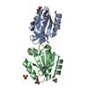
| Deposited unit | 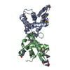
| |||||||||||||||||||||||||||||||||||||||
|---|---|---|---|---|---|---|---|---|---|---|---|---|---|---|---|---|---|---|---|---|---|---|---|---|---|---|---|---|---|---|---|---|---|---|---|---|---|---|---|---|
| 1 |
| |||||||||||||||||||||||||||||||||||||||
| Unit cell |
| |||||||||||||||||||||||||||||||||||||||
| Noncrystallographic symmetry (NCS) | NCS domain:
NCS domain segments:
| |||||||||||||||||||||||||||||||||||||||
| Details | CRYSTAL PACKING ANALYSIS SUGGESTS THE ASSIGNMENT OF A DIMER AS THE SIGNIFICANT OLIGOMERIZATION STATE. |
-Components
| #1: Protein | Mass: 14504.097 Da / Num. of mol.: 2 Source method: isolated from a genetically manipulated source Source: (gene. exp.) Homo sapiens (human) / Gene: BC063280, INO80G, NFRKB / Plasmid: SpeedET / Production host:  Escherichia Coli (E. coli) / Strain (production host): HK100 / References: UniProt: Q6P4R8 Escherichia Coli (E. coli) / Strain (production host): HK100 / References: UniProt: Q6P4R8#2: Chemical | ChemComp-NA / |   Mass: 22.990 Da / Num. of mol.: 1 / Source method: obtained synthetically / Formula: Na Mass: 22.990 Da / Num. of mol.: 1 / Source method: obtained synthetically / Formula: Na#3: Water | ChemComp-HOH / | Water Mass: 18.015 Da / Num. of mol.: 25 / Source method: isolated from a natural source / Formula: H2O Mass: 18.015 Da / Num. of mol.: 25 / Source method: isolated from a natural source / Formula: H2OSequence details | 1. THIS CONSTRUCT (RESIDUES 370-495) WAS EXPRESSED WITH AN N-TERMINAL PURIFICATION TAG ...1. THIS CONSTRUCT (RESIDUES 370-495) WAS EXPRESSED WITH AN N-TERMINAL PURIFICATI | |
|---|
-Experimental details
-Experiment
| Experiment | Method: X-RAY DIFFRACTION / Number of used crystals: 1 |
|---|
- Sample preparation
| Crystal | Density Matthews: 2.09 Å3/Da / Density % sol: 41.26 % |
|---|---|
| Crystal grow | Temperature: 277 K / Method: vapor diffusion, sitting drop / pH: 7.5 Details: 0.09M HEPES pH 7.5, 10% glycerol, 1.26M tri-sodium citrate, NANODROP, VAPOR DIFFUSION, SITTING DROP, temperature 277K |
-Data collection
| Diffraction | Mean temperature: 100 K | |||||||||||||||||||||||||||||||||||||||||||||||||||||||||||||||||||||||||||||
|---|---|---|---|---|---|---|---|---|---|---|---|---|---|---|---|---|---|---|---|---|---|---|---|---|---|---|---|---|---|---|---|---|---|---|---|---|---|---|---|---|---|---|---|---|---|---|---|---|---|---|---|---|---|---|---|---|---|---|---|---|---|---|---|---|---|---|---|---|---|---|---|---|---|---|---|---|---|---|
| Diffraction source | Source: SYNCHROTRON / Site: SSRL  / Beamline: BL9-2 / Wavelength: 0.91837,0.97936,0.97915 / Beamline: BL9-2 / Wavelength: 0.91837,0.97936,0.97915 | |||||||||||||||||||||||||||||||||||||||||||||||||||||||||||||||||||||||||||||
| Detector | Type: MARMOSAIC 325 mm CCD / Detector: CCD / Date: Jun 22, 2011 / Details: double crystal monochromator | |||||||||||||||||||||||||||||||||||||||||||||||||||||||||||||||||||||||||||||
| Radiation | Monochromator: double crystal / Protocol: MAD / Monochromatic (M) / Laue (L): M / Scattering type: x-ray | |||||||||||||||||||||||||||||||||||||||||||||||||||||||||||||||||||||||||||||
| Radiation wavelength |
| |||||||||||||||||||||||||||||||||||||||||||||||||||||||||||||||||||||||||||||
| Reflection | Resolution: 2.18→35.466 Å / Num. obs: 13560 / % possible obs: 99.8 % / Observed criterion σ(I): -3 / Biso Wilson estimate: 41.259 Å2 / Rmerge(I) obs: 0.067 / Net I/σ(I): 20.78 | |||||||||||||||||||||||||||||||||||||||||||||||||||||||||||||||||||||||||||||
| Reflection shell | Diffraction-ID: 1
|
-Phasing
| Phasing | Method: MAD |
|---|
- Processing
| Software |
| |||||||||||||||||||||||||||||||||||||||||||||||||||||||||||||||||||||||||||||||||||||
|---|---|---|---|---|---|---|---|---|---|---|---|---|---|---|---|---|---|---|---|---|---|---|---|---|---|---|---|---|---|---|---|---|---|---|---|---|---|---|---|---|---|---|---|---|---|---|---|---|---|---|---|---|---|---|---|---|---|---|---|---|---|---|---|---|---|---|---|---|---|---|---|---|---|---|---|---|---|---|---|---|---|---|---|---|---|---|
| Refinement | Method to determine structure: MAD / Resolution: 2.18→35.466 Å / Cor.coef. Fo:Fc: 0.929 / Cor.coef. Fo:Fc free: 0.905 / Occupancy max: 1 / Occupancy min: 0.37 / SU B: 12.518 / SU ML: 0.156 / Cross valid method: THROUGHOUT / σ(F): 0 / ESU R Free: 0.222 Stereochemistry target values: MAXIMUM LIKELIHOOD WITH PHASES Details: 1. HYDROGENS HAVE BEEN ADDED IN THE RIDING POSITIONS. 2. ATOM RECORD CONTAINS SUM OF TLS AND RESIDUAL B FACTORS. 3. ANISOU RECORD CONTAINS SUM OF TLS AND RESIDUAL U FACTORS. 4. WATERS WERE ...Details: 1. HYDROGENS HAVE BEEN ADDED IN THE RIDING POSITIONS. 2. ATOM RECORD CONTAINS SUM OF TLS AND RESIDUAL B FACTORS. 3. ANISOU RECORD CONTAINS SUM OF TLS AND RESIDUAL U FACTORS. 4. WATERS WERE EXCLUDED FROM AUTOMATIC TLS ASSIGNMENT. 5. A MET-INHIBITION PROTOCOL WAS USED FOR SELENOMETHIONINE INCORPORATION DURING PROTEIN EXPRESSION. THE OCCUPANCY OF THE SE ATOMS IN THE MSE RESIDUES WAS REDUCED TO 0.75 FOR THE REDUCED SCATTERING POWER DUE TO PARTIAL S-MET INCORPORATION. 6. SODIUM ION (NA) FROM CRYSTALLIZATION CONDITION WAS MODELED.
| |||||||||||||||||||||||||||||||||||||||||||||||||||||||||||||||||||||||||||||||||||||
| Solvent computation | Ion probe radii: 0.8 Å / Shrinkage radii: 0.8 Å / VDW probe radii: 1.2 Å / Solvent model: BABINET MODEL WITH MASK | |||||||||||||||||||||||||||||||||||||||||||||||||||||||||||||||||||||||||||||||||||||
| Displacement parameters | Biso max: 97.07 Å2 / Biso mean: 46.2329 Å2 / Biso min: 25.43 Å2
| |||||||||||||||||||||||||||||||||||||||||||||||||||||||||||||||||||||||||||||||||||||
| Refinement step | Cycle: LAST / Resolution: 2.18→35.466 Å
| |||||||||||||||||||||||||||||||||||||||||||||||||||||||||||||||||||||||||||||||||||||
| Refine LS restraints |
| |||||||||||||||||||||||||||||||||||||||||||||||||||||||||||||||||||||||||||||||||||||
| Refine LS restraints NCS | Dom-ID: 1 / Auth asym-ID: A / Ens-ID: 1 / Number: 1360 / Refine-ID: X-RAY DIFFRACTION
| |||||||||||||||||||||||||||||||||||||||||||||||||||||||||||||||||||||||||||||||||||||
| LS refinement shell | Resolution: 2.18→2.236 Å / Total num. of bins used: 20
| |||||||||||||||||||||||||||||||||||||||||||||||||||||||||||||||||||||||||||||||||||||
| Refinement TLS params. | Method: refined / Refine-ID: X-RAY DIFFRACTION
| |||||||||||||||||||||||||||||||||||||||||||||||||||||||||||||||||||||||||||||||||||||
| Refinement TLS group |
|
