- PDB-3txn: Crystal structure of Rpn6 from Drosophila melanogaster, native data -
+
Open data
ID or keywords:
Loading...
-
Basic information
Entry
Database: PDB / ID: 3txn
Title
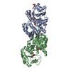
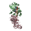
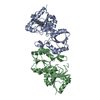
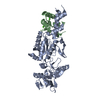
Crystal structure of Rpn6 from Drosophila melanogaster, native data
Components
26S proteasome regulatory complex subunit p42B
Keywords
HYDROLASE / PROTEIN BINDING / 26 S proteasome / PCI domain / alpha solenoid / regulatory particle / lid
Function / homology
Function and homology information
Activation of NF-kappaB in B cells / Cross-presentation of soluble exogenous antigens (endosomes) / Downstream TCR signaling / Degradation of NF-kappa-B inhibitor, CACT / FCERI mediated NF-kB activation / Regulation of ornithine decarboxylase (ODC) / CLEC7A (Dectin-1) signaling / AUF1 (hnRNP D0) binds and destabilizes mRNA / Circadian Clock pathway / Oxygen-dependent proline hydroxylation of Hypoxia-inducible Factor Alpha ...Activation of NF-kappaB in B cells / Cross-presentation of soluble exogenous antigens (endosomes) / Downstream TCR signaling / Degradation of NF-kappa-B inhibitor, CACT / FCERI mediated NF-kB activation / Regulation of ornithine decarboxylase (ODC) / CLEC7A (Dectin-1) signaling / AUF1 (hnRNP D0) binds and destabilizes mRNA / Circadian Clock pathway / Oxygen-dependent proline hydroxylation of Hypoxia-inducible Factor Alpha / Autodegradation of Cdh1 by Cdh1:APC/C / APC/C:Cdc20 mediated degradation of Securin / APC/C:Cdh1 mediated degradation of Cdc20 and other APC/C:Cdh1 targeted proteins in late mitosis/early G1 / SCF(Skp2)-mediated degradation of p27/p21 / Degradation of beta-catenin by the destruction complex / Ubiquitination and degradation of phosphorylated ARM / Separation of Sister Chromatids / Degradation of AXIN / Degradation of DVL / Dectin-1 mediated noncanonical NF-kB signaling / Regulation of RAS by GAPs / NIK-->noncanonical NF-kB signaling / Ubiquitin Mediated Degradation of Phosphorylated Cdc25A / Ubiquitin-dependent degradation of Cyclin D / FBXL7 down-regulates AURKA during mitotic entry and in early mitosis / Regulation of RUNX2 expression and activity / Regulation of RUNX3 expression and activity / KEAP1-NFE2L2 pathway / GSK3B and BTRC:CUL1-mediated-degradation of NFE2L2 / Nuclear CI is degraded / ABC-family proteins mediated transport / Cdc20:Phospho-APC/C mediated degradation of Cyclin A / Ubiquitination and proteolysis of phosphorylated CI / RUNX1 regulates transcription of genes involved in differentiation of HSCs / Interleukin-1 signaling / Degradation of GLI1 by the proteasome / GLI3 is processed to GLI3R by the proteasome / Hedgehog 'on' state / Assembly of the pre-replicative complex / Hedgehog ligand biogenesis / Degradation of PER / Asymmetric localization of PCP proteins / Degradation of CRY / CDK-mediated phosphorylation and removal of Cdc6 / Neddylation / Regulation of PTEN stability and activity / Degradation of TIM / Antigen processing: Ubiquitination & Proteasome degradation / Orc1 removal from chromatin / Ub-specific processing proteases / UCH proteinases / Neutrophil degranulation / proteasome regulatory particle / proteasome regulatory particle, lid subcomplex / proteasome complex / ubiquitin-dependent protein catabolic process / proteasome-mediated ubiquitin-dependent protein catabolic process / structural molecule activity / nucleoplasm / cytosol Similarity search - Function
In the structure databanks used in Yorodumi, some data are registered as the other names, "COVID-19 virus" and "2019-nCoV". Here are the details of the virus and the list of structure data.
Jan 31, 2019. EMDB accession codes are about to change! (news from PDBe EMDB page)
EMDB accession codes are about to change! (news from PDBe EMDB page)
The allocation of 4 digits for EMDB accession codes will soon come to an end. Whilst these codes will remain in use, new EMDB accession codes will include an additional digit and will expand incrementally as the available range of codes is exhausted. The current 4-digit format prefixed with “EMD-” (i.e. EMD-XXXX) will advance to a 5-digit format (i.e. EMD-XXXXX), and so on. It is currently estimated that the 4-digit codes will be depleted around Spring 2019, at which point the 5-digit format will come into force.
The EM Navigator/Yorodumi systems omit the EMD- prefix.
Related info.:Q: What is EMD? / ID/Accession-code notation in Yorodumi/EM Navigator
Yorodumi is a browser for structure data from EMDB, PDB, SASBDB, etc.
This page is also the successor to EM Navigator detail page, and also detail information page/front-end page for Omokage search.
The word "yorodu" (or yorozu) is an old Japanese word meaning "ten thousand". "mi" (miru) is to see.
Related info.:EMDB / PDB / SASBDB / Comparison of 3 databanks / Yorodumi Search / Aug 31, 2016. New EM Navigator & Yorodumi / Yorodumi Papers / Jmol/JSmol / Function and homology information / Changes in new EM Navigator and Yorodumi
 Movie
Movie Controller

 Yorodumi
 Open data
 Basic information
 Components
 Keywords
 HYDROLASE /
  Function and homology information
 Authors
 Citation
 Structure visualization
 Downloads & links
 Other downloads


 PDBj
PDBj


 Assembly


 Mass: 92.094 Da / Num. of mol.: 2 / Source method: obtained synthetically / Formula: C3H8O3
 Mass: 96.063 Da / Num. of mol.: 2 / Source method: obtained synthetically / Formula: SO4
 Mass: 18.015 Da / Num. of mol.: 49 / Source method: isolated from a natural source / Formula: H2O
 Sample preparation
 / Beamline: ID29 / Wavelength: 1.00686 Å
 Processing