 Movie
Movie Controller

[English] 日本語
 Yorodumi
- PDB-3tw9: Crystal structure of gluconate dehydratase (TARGET EFI-501679) fr... -
+ Open data

- Basic information
| Entry | Database: PDB / ID: 3tw9 | ||||||
|---|---|---|---|---|---|---|---|
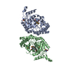
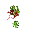
| Title | Crystal structure of gluconate dehydratase (TARGET EFI-501679) from Salmonella enterica subsp. enterica serovar Enteritidis str. P125109 | ||||||
 Components Components | Putative dehydratase | ||||||
 Keywords Keywords | LYASE / ENOLASE / MAGNESIUM BINDING SITE / Structural Genomics | ||||||
| Function / homology |  Function and homology informationgluconate dehydratase / gluconate dehydratase activity / Lyases; Carbon-oxygen lyases; Hydro-lyases / amino acid catabolic process / carbohydrate catabolic process / magnesium ion binding Function and homology informationgluconate dehydratase / gluconate dehydratase activity / Lyases; Carbon-oxygen lyases; Hydro-lyases / amino acid catabolic process / carbohydrate catabolic process / magnesium ion bindingSimilarity search - Function | ||||||
| Biological species |  Salmonella enterica subsp. enterica serovar Enteritidis (bacteria) Salmonella enterica subsp. enterica serovar Enteritidis (bacteria) | ||||||
| Method | X-RAY DIFFRACTION / SYNCHROTRON / MOLECULAR REPLACEMENT / Resolution: 1.7 Å | ||||||
 Authors Authors | Patskovsky, Y. / Toro, R. / Bhosle, R. / Hillerich, B. / Seidel, R.D. / Washington, E. / Scott Glenn, A. / Chowdhury, S. / Evans, B. / Hammonds, J. ...Patskovsky, Y. / Toro, R. / Bhosle, R. / Hillerich, B. / Seidel, R.D. / Washington, E. / Scott Glenn, A. / Chowdhury, S. / Evans, B. / Hammonds, J. / Zencheck, W.D. / Imker, H.J. / Gerlt, J.A. / Almo, S.C. / Enzyme Function Initiative (EFI) | ||||||
 Citation Citation | Journal: To be Published Title: Crystal Structure of Gluconate Dehydratase from Salmonella Enterica P125109 Authors: Patskovsky, Y. / Toro, R. / Bhosle, R. / Hillerich, B. / Seidel, R.D. / Washington, E. / Scott Glenn, A. / Chowdhury, S. / Evans, B. / Hammonds, J. / Zencheck, W.D. / Imker, H.J. / Gerlt, J.A. / Almo, S.C. | ||||||
| History |
|
- Structure visualization
| Structure viewer | Molecule: MolmilJmol/JSmol |
|---|
- Downloads & links
-Download
| PDBx/mmCIF format | 3tw9.cif.gz | 347.3 KB | Display | PDBx/mmCIF format |
|---|---|---|---|---|
| PDB format | pdb3tw9.ent.gz | 279.7 KB | Display | PDB format |
| PDBx/mmJSON format | 3tw9.json.gz | Tree view | PDBx/mmJSON format | |
| Others |  Other downloads Other downloads |
-Validation report
| Arichive directory | https://data.pdbj.org/pub/pdb/validation_reports/tw/3tw9ftp://data.pdbj.org/pub/pdb/validation_reports/tw/3tw9 | HTTPS FTP |
|---|
-Related structure data
| Related structure data | 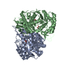 3twaC 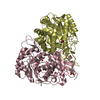 3twbC 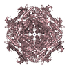 3dfhS C: citing same article ( S: Starting model for refinement |
|---|---|
| Similar structure data | |
| Other databases |


-Links
 PDBj
PDBj
- Assembly
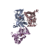
| Deposited unit | 
| |||||||||||||||||||||
|---|---|---|---|---|---|---|---|---|---|---|---|---|---|---|---|---|---|---|---|---|---|---|
| 1 | 
| |||||||||||||||||||||
| 2 | 
| |||||||||||||||||||||
| 3 | 
| |||||||||||||||||||||
| Unit cell |
| |||||||||||||||||||||
| Components on special symmetry positions |
|
-Components
| #1: Protein | Mass: 49042.746 Da / Num. of mol.: 4 Source method: isolated from a genetically manipulated source Source: (gene. exp.) Salmonella enterica subsp. enterica serovar Enteritidis (bacteria)Strain: P125109 / Gene: SEN1436 / Plasmid: PET / Production host: Escherichia coli (E. coli) / Strain (production host): BL21(DE3) / References: UniProt: B5R541#2: Chemical | ChemComp-GOL / Glycerol  Mass: 92.094 Da / Num. of mol.: 4 / Source method: obtained synthetically / Formula: C3H8O3 Mass: 92.094 Da / Num. of mol.: 4 / Source method: obtained synthetically / Formula: C3H8O3#3: Chemical | ChemComp-CL / Chloride  Mass: 35.453 Da / Num. of mol.: 11 / Source method: obtained synthetically / Formula: Cl Mass: 35.453 Da / Num. of mol.: 11 / Source method: obtained synthetically / Formula: Cl#4: Water | ChemComp-HOH / | Water Mass: 18.015 Da / Num. of mol.: 1171 / Source method: isolated from a natural source / Formula: H2O Mass: 18.015 Da / Num. of mol.: 1171 / Source method: isolated from a natural source / Formula: H2O |
|---|
-Experimental details
-Experiment
| Experiment | Method: X-RAY DIFFRACTION / Number of used crystals: 1 |
|---|
- Sample preparation
| Crystal | Density Matthews: 2.21 Å3/Da / Density % sol: 44.48 % |
|---|---|
| Crystal grow | Method: vapor diffusion, sitting drop / pH: 4.6 Details: 0.2M SODIUM CHLORIDE, 100MM SODIUM ACETATE, PH 4.6, 30% MPD, VAPOR DIFFUSION, SITTING DROP, TEMPERATURE 294K |
-Data collection
| Diffraction | Mean temperature: 100 K |
|---|---|
| Diffraction source | Source: SYNCHROTRON / Site: NSLS  / Beamline: X29A / Wavelength: 1.075 / Beamline: X29A / Wavelength: 1.075 |
| Detector | Type: ADSC QUANTUM 315 / Detector: CCD / Date: Jul 23, 2011 / Details: MIRRORS |
| Radiation | Protocol: SINGLE WAVELENGTH / Monochromatic (M) / Laue (L): M / Scattering type: x-ray |
| Radiation wavelength | Wavelength: 1.075 Å / Relative weight: 1 |
| Reflection | Resolution: 1.65→50 Å / Num. obs: 209416 / % possible obs: 100 % / Observed criterion σ(I): -5 / Redundancy: 7.6 % / Biso Wilson estimate: 21.13 Å2 / Rsym value: 0.129 / Net I/σ(I): 5.2 |
| Reflection shell | Resolution: 1.65→1.68 Å / Redundancy: 7.3 % / Rmerge(I) obs: 0.96 / Mean I/σ(I) obs: 1.3 / % possible all: 100 |
- Processing
| Software |
| ||||||||||||||||||||||||||||||||||||||||||||||||||||||||||||||||||||||||||||||||||||||||||||||||||||||||||||||||||||||||||||||||||||||||||||||||||||||||||||||||||||||||||
|---|---|---|---|---|---|---|---|---|---|---|---|---|---|---|---|---|---|---|---|---|---|---|---|---|---|---|---|---|---|---|---|---|---|---|---|---|---|---|---|---|---|---|---|---|---|---|---|---|---|---|---|---|---|---|---|---|---|---|---|---|---|---|---|---|---|---|---|---|---|---|---|---|---|---|---|---|---|---|---|---|---|---|---|---|---|---|---|---|---|---|---|---|---|---|---|---|---|---|---|---|---|---|---|---|---|---|---|---|---|---|---|---|---|---|---|---|---|---|---|---|---|---|---|---|---|---|---|---|---|---|---|---|---|---|---|---|---|---|---|---|---|---|---|---|---|---|---|---|---|---|---|---|---|---|---|---|---|---|---|---|---|---|---|---|---|---|---|---|---|---|---|
| Refinement | Method to determine structure: MOLECULAR REPLACEMENT Starting model: PDB ENTRY 3DFH Resolution: 1.7→50 Å / Cor.coef. Fo:Fc: 0.965 / Cor.coef. Fo:Fc free: 0.951 / SU B: 2.573 / SU ML: 0.084 / Cross valid method: THROUGHOUT / ESU R: 0.119 / ESU R Free: 0.118 / Stereochemistry target values: MAXIMUM LIKELIHOOD / Details: HYDROGENS HAVE BEEN ADDED IN THE RIDING POSITIONS
| ||||||||||||||||||||||||||||||||||||||||||||||||||||||||||||||||||||||||||||||||||||||||||||||||||||||||||||||||||||||||||||||||||||||||||||||||||||||||||||||||||||||||||
| Solvent computation | Ion probe radii: 0.8 Å / Shrinkage radii: 0.8 Å / VDW probe radii: 1.4 Å / Solvent model: BABINET MODEL WITH MASK | ||||||||||||||||||||||||||||||||||||||||||||||||||||||||||||||||||||||||||||||||||||||||||||||||||||||||||||||||||||||||||||||||||||||||||||||||||||||||||||||||||||||||||
| Displacement parameters | Biso mean: 29.093 Å2
| ||||||||||||||||||||||||||||||||||||||||||||||||||||||||||||||||||||||||||||||||||||||||||||||||||||||||||||||||||||||||||||||||||||||||||||||||||||||||||||||||||||||||||
| Refinement step | Cycle: LAST / Resolution: 1.7→50 Å
| ||||||||||||||||||||||||||||||||||||||||||||||||||||||||||||||||||||||||||||||||||||||||||||||||||||||||||||||||||||||||||||||||||||||||||||||||||||||||||||||||||||||||||
| Refine LS restraints |
| ||||||||||||||||||||||||||||||||||||||||||||||||||||||||||||||||||||||||||||||||||||||||||||||||||||||||||||||||||||||||||||||||||||||||||||||||||||||||||||||||||||||||||
| LS refinement shell | Resolution: 1.7→1.744 Å / Total num. of bins used: 20
|
