 Movie
Movie Controller

[English] 日本語
 Yorodumi
- PDB-3tw0: Structural Analysis of Adhesive Tip pilin, GBS104 from Group B St... -
+ Open data

- Basic information
| Entry | Database: PDB / ID: 3tw0 | ||||||
|---|---|---|---|---|---|---|---|
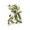
| Title | Structural Analysis of Adhesive Tip pilin, GBS104 from Group B Streptococcus agalactiae | ||||||
 Components Components | Cell wall surface anchor family protein | ||||||
 Keywords Keywords |  CELL ADHESION / vWFA fold / Ancillary pilin / Gram-positive bacterial cell surface CELL ADHESION / vWFA fold / Ancillary pilin / Gram-positive bacterial cell surface | ||||||
| Function / homology |  Function and homology information Function and homology information | ||||||
| Biological species |  Streptococcus agalactiae serogroup V (bacteria) Streptococcus agalactiae serogroup V (bacteria) | ||||||
| Method | X-RAY DIFFRACTION / MOLECULAR REPLACEMENT / Resolution: 2 Å | ||||||
 Authors Authors | Krishnan, V. / Narayana, S.V.L. | ||||||
 Citation Citation | Journal: Acta Crystallogr.,Sect.D / Year: 2013 Title: Structure of Streptococcus agalactiae tip pilin GBS104: a model for GBS pili assembly and host interactions. Authors: Krishnan, V. / Dwivedi, P. / Kim, B.J. / Samal, A. / Macon, K. / Ma, X. / Mishra, A. / Doran, K.S. / Ton-That, H. / Narayana, S.V. | ||||||
| History |
|
- Structure visualization
| Structure viewer | Molecule: MolmilJmol/JSmol |
|---|
- Downloads & links
-Download
| PDBx/mmCIF format | 3tw0.cif.gz | 304.2 KB | Display | PDBx/mmCIF format |
|---|---|---|---|---|
| PDB format | pdb3tw0.ent.gz | 245.5 KB | Display | PDB format |
| PDBx/mmJSON format | 3tw0.json.gz | Tree view | PDBx/mmJSON format | |
| Others |  Other downloads Other downloads |
-Validation report
| Arichive directory | https://data.pdbj.org/pub/pdb/validation_reports/tw/3tw0ftp://data.pdbj.org/pub/pdb/validation_reports/tw/3tw0 | HTTPS FTP |
|---|
-Related structure data
| Related structure data |  3tvySC  3txaC S: Starting model for refinement C: citing same article ( |
|---|---|
| Similar structure data |


-Links
 PDBj
PDBj


- Assembly
| Deposited unit | 
| ||||||||
|---|---|---|---|---|---|---|---|---|---|
| 1 | 
| ||||||||
| 2 | 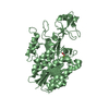
| ||||||||
| 3 | 
| ||||||||
| 4 | 
| ||||||||
| Unit cell |
|
-Components
| #1: Protein | Mass: 41639.133 Da / Num. of mol.: 4 / Fragment: SEE REMARK 999 / Mutation: T564C, K571C Source method: isolated from a genetically manipulated source Source: (gene. exp.) Streptococcus agalactiae serogroup V (bacteria)Strain: 2603 V/R / Gene: SAG0649 / Plasmid: pMCSG7 / Production host: Escherichia coli (E. coli) / Strain (production host): BL21(DE3) / References: UniProt: Q8E0S5#2: Chemical | ChemComp-MG /   Mass: 24.305 Da / Num. of mol.: 4 / Source method: obtained synthetically / Formula: Mg Mass: 24.305 Da / Num. of mol.: 4 / Source method: obtained synthetically / Formula: Mg#3: Chemical | ChemComp-ACT / Acetate  Mass: 59.044 Da / Num. of mol.: 4 / Source method: obtained synthetically / Formula: C2H3O2 Mass: 59.044 Da / Num. of mol.: 4 / Source method: obtained synthetically / Formula: C2H3O2#4: Water | ChemComp-HOH / | Water Mass: 18.015 Da / Num. of mol.: 759 / Source method: isolated from a natural source / Formula: H2O Mass: 18.015 Da / Num. of mol.: 759 / Source method: isolated from a natural source / Formula: H2OSequence details | N3 DOMAIN (UNP RESIDUES 212-581) REMAINS AFTER DEGRADATION OF ORIGINAL PROTEIN CONSTRUCT (UNP ...N3 DOMAIN (UNP RESIDUES 212-581) REMAINS AFTER DEGRADATIO | |
|---|
-Experimental details
-Experiment
| Experiment | Method: X-RAY DIFFRACTION / Number of used crystals: 1 |
|---|
- Sample preparation
| Crystal | Density Matthews: 2.14 Å3/Da / Density % sol: 42.49 % |
|---|---|
| Crystal grow | Temperature: 295 K / Method: vapor diffusion, hanging drop / pH: 7.5 Details: 20% PEG3000, 0.1 M HEPES, 0.2 M NaCl, 10 mM Spermine-4HCl, pH 7.5, VAPOR DIFFUSION, HANGING DROP, temperature 295K |
-Data collection
| Diffraction | Mean temperature: 100 K |
|---|---|
| Diffraction source | Source: ROTATING ANODE / Type: RIGAKU / Wavelength: 1.5418 Å |
| Detector | Type: RIGAKU RAXIS IV / Detector: IMAGE PLATE / Date: Apr 14, 2010 |
| Radiation | Protocol: SINGLE WAVELENGTH / Monochromatic (M) / Laue (L): M / Scattering type: x-ray |
| Radiation wavelength | Wavelength: 1.5418 Å / Relative weight: 1 |
| Reflection | Resolution: 2→46.304 Å / Num. obs: 88454 / % possible obs: 89 % / Redundancy: 3.6 % / Rmerge(I) obs: 0.07 / Net I/σ(I): 11.5 |
| Reflection shell | Resolution: 2→2.07 Å / Redundancy: 3.7 % / Rmerge(I) obs: 0.29 / Mean I/σ(I) obs: 4.2 / % possible all: 80.2 |
- Processing
| Software |
| |||||||||||||||||||||||||||||||||||||||||||||||||||||||||||||||||
|---|---|---|---|---|---|---|---|---|---|---|---|---|---|---|---|---|---|---|---|---|---|---|---|---|---|---|---|---|---|---|---|---|---|---|---|---|---|---|---|---|---|---|---|---|---|---|---|---|---|---|---|---|---|---|---|---|---|---|---|---|---|---|---|---|---|---|
| Refinement | Method to determine structure: MOLECULAR REPLACEMENT Starting model: PDB ENTRY 3TVY Resolution: 2→46.304 Å / Cor.coef. Fo:Fc: 0.94 / Cor.coef. Fo:Fc free: 0.922 / SU B: 3.843 / SU ML: 0.107 / Cross valid method: THROUGHOUT / ESU R: 0.246 / ESU R Free: 0.186 / Stereochemistry target values: MAXIMUM LIKELIHOOD Details: HYDROGENS HAVE BEEN ADDED IN THE RIDING POSITIONS U VALUES : REFINED INDIVIDUALLY
| |||||||||||||||||||||||||||||||||||||||||||||||||||||||||||||||||
| Solvent computation | Ion probe radii: 0.8 Å / Shrinkage radii: 0.8 Å / VDW probe radii: 1.4 Å / Solvent model: MASK | |||||||||||||||||||||||||||||||||||||||||||||||||||||||||||||||||
| Displacement parameters | Biso mean: 24.932 Å2
| |||||||||||||||||||||||||||||||||||||||||||||||||||||||||||||||||
| Refinement step | Cycle: LAST / Resolution: 2→46.304 Å
| |||||||||||||||||||||||||||||||||||||||||||||||||||||||||||||||||
| Refine LS restraints |
| |||||||||||||||||||||||||||||||||||||||||||||||||||||||||||||||||
| LS refinement shell | Resolution: 2→2.052 Å / Total num. of bins used: 20
|
