 Movie
Movie Controller

[English] 日本語
 Yorodumi
- PDB-3tul: Crystal structure of N-terminal region of Type III Secretion Majo... -
+ Open data

- Basic information
| Entry | Database: PDB / ID: 3tul | ||||||
|---|---|---|---|---|---|---|---|
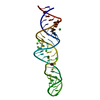
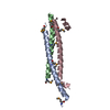
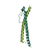
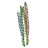
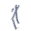
| Title | Crystal structure of N-terminal region of Type III Secretion Major Translocator SipB (residues 82-226) | ||||||
 Components Components | Cell invasion protein sipB | ||||||
 Keywords Keywords | CELL INVASION / translocator /  type three secretion system / coiled-coil / virulence type three secretion system / coiled-coil / virulence | ||||||
| Function / homology |  Function and homology information Function and homology informationprotein localization to Golgi apparatus / : / membrane => GO:0016020 / host cell plasma membrane / extracellular region Similarity search - Function | ||||||
| Biological species |  Salmonella enterica subsp. enterica serovar Typhimurium (bacteria) Salmonella enterica subsp. enterica serovar Typhimurium (bacteria) | ||||||
| Method | X-RAY DIFFRACTION / SYNCHROTRON / MAD / Resolution: 2.793 Å | ||||||
 Authors Authors | Barta, M.L. / Dickenson, N.E. / Patel, M. / Keightley, J.A. / Picking, W.D. / Picking, W.L. / Geisbrecht, B.V. | ||||||
 Citation Citation | Journal: J.Mol.Biol. / Year: 2012 Title: The Structures of Coiled-Coil Domains from Type III Secretion System Translocators Reveal Homology to Pore-Forming Toxins. Authors: Barta, M.L. / Dickenson, N.E. / Patil, M. / Keightley, A. / Wyckoff, G.J. / Picking, W.D. / Picking, W.L. / Geisbrecht, B.V. | ||||||
| History |
|
- Structure visualization
| Structure viewer | Molecule: MolmilJmol/JSmol |
|---|
- Downloads & links
-Download
| PDBx/mmCIF format | 3tul.cif.gz | 198.1 KB | Display | PDBx/mmCIF format |
|---|---|---|---|---|
| PDB format | pdb3tul.ent.gz | 168 KB | Display | PDB format |
| PDBx/mmJSON format | 3tul.json.gz | Tree view | PDBx/mmJSON format | |
| Others |  Other downloads Other downloads |
-Validation report
| Arichive directory | https://data.pdbj.org/pub/pdb/validation_reports/tu/3tulftp://data.pdbj.org/pub/pdb/validation_reports/tu/3tul | HTTPS FTP |
|---|
-Related structure data


-Links
 PDBj
PDBj- Assembly
| Deposited unit | 
| ||||||||
|---|---|---|---|---|---|---|---|---|---|
| 1 | 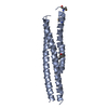
| ||||||||
| 2 | 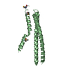
| ||||||||
| 3 | 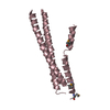
| ||||||||
| 4 | 
| ||||||||
| 5 |
| ||||||||
| Unit cell |
|
-Components
| #1: Protein | Mass: 16838.328 Da / Num. of mol.: 4 / Fragment: N-terminal domain (UNP residues 81-237) Source method: isolated from a genetically manipulated source Source: (gene. exp.) Salmonella enterica subsp. enterica serovar Typhimurium (bacteria)Gene: sipB, sspB, STM2885 / Plasmid: pT7HMT / Production host: Escherichia coli (E. coli) / Strain (production host): BL21(DE3) / References: UniProt: Q56019#2: Water | ChemComp-HOH / | Water Mass: 18.015 Da / Num. of mol.: 43 / Source method: isolated from a natural source / Formula: H2O Mass: 18.015 Da / Num. of mol.: 43 / Source method: isolated from a natural source / Formula: H2O |
|---|
-Experimental details
-Experiment
| Experiment | Method: X-RAY DIFFRACTION / Number of used crystals: 1 |
|---|
- Sample preparation
| Crystal | Density Matthews: 2.56 Å3/Da / Density % sol: 51.9 % |
|---|---|
| Crystal grow | Temperature: 293 K / Method: vapor diffusion, hanging drop / pH: 7.1 Details: 0.15 M potassium bromide, 27% PEG2000 MME, pH 7.1, VAPOR DIFFUSION, HANGING DROP, temperature 293K |
-Data collection
| Diffraction | Mean temperature: 100 K | |||||||||
|---|---|---|---|---|---|---|---|---|---|---|
| Diffraction source | Source: SYNCHROTRON / Site: APS  / Beamline: 22-BM / Wavelength: 0.97243,0.97934 / Beamline: 22-BM / Wavelength: 0.97243,0.97934 | |||||||||
| Detector | Type: MARMOSAIC 225 mm CCD / Detector: CCD / Date: Jul 16, 2011 / Details: mirror | |||||||||
| Radiation | Monochromator: Si(220) / Protocol: MAD / Monochromatic (M) / Laue (L): M / Scattering type: x-ray | |||||||||
| Radiation wavelength |
| |||||||||
| Reflection | Resolution: 2.793→31.943 Å / Num. all: 17834 / Num. obs: 17478 / % possible obs: 98 % / Observed criterion σ(F): 0 / Observed criterion σ(I): 2 / Redundancy: 12 % / Rmerge(I) obs: 0.1 / Net I/σ(I): 18.8 | |||||||||
| Reflection shell | Resolution: 2.793→2.9681 Å / Rmerge(I) obs: 0.671 / Mean I/σ(I) obs: 2.55 / % possible all: 80 |
- Processing
| Software |
| |||||||||||||||||||||||||||||||||||||||||||||||||
|---|---|---|---|---|---|---|---|---|---|---|---|---|---|---|---|---|---|---|---|---|---|---|---|---|---|---|---|---|---|---|---|---|---|---|---|---|---|---|---|---|---|---|---|---|---|---|---|---|---|---|
| Refinement | Method to determine structure: MAD / Resolution: 2.793→31.943 Å / Occupancy max: 1 / Occupancy min: 1 / SU ML: 0.39 / σ(F): 0.26 / Phase error: 34.67 / Stereochemistry target values: MLHL
| |||||||||||||||||||||||||||||||||||||||||||||||||
| Solvent computation | Shrinkage radii: 0.72 Å / VDW probe radii: 1 Å / Solvent model: FLAT BULK SOLVENT MODEL / Bsol: 26.993 Å2 / ksol: 0.266 e/Å3 | |||||||||||||||||||||||||||||||||||||||||||||||||
| Displacement parameters |
| |||||||||||||||||||||||||||||||||||||||||||||||||
| Refinement step | Cycle: LAST / Resolution: 2.793→31.943 Å
| |||||||||||||||||||||||||||||||||||||||||||||||||
| Refine LS restraints |
| |||||||||||||||||||||||||||||||||||||||||||||||||
| LS refinement shell |
|
