 Movie
Movie Controller

[English] 日本語
 Yorodumi
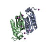
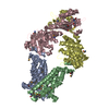
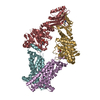
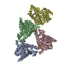
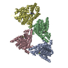
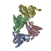
- PDB-3tui: Inward facing conformations of the MetNI methionine ABC transport... -
+ Open data

- Basic information
| Entry | Database: PDB / ID: 3tui | ||||||
|---|---|---|---|---|---|---|---|
| Title | Inward facing conformations of the MetNI methionine ABC transporter: CY5 native crystal form | ||||||
 Components Components |
| ||||||
 Keywords Keywords | HYDROLASE/TRANSPORT PROTEIN /  ABC-TRANSPORTER / TYPE I ABC TYPE IMPORTER / METHIONINE UPTAKE TRANSPORTER / MEMBRANE PROTEIN / AMINO-ACID TRANSPORT / ATP-BINDING / HYDROLASE / INNER MEMBRANE / HYDROLASE-TRANSPORT PROTEIN complex ABC-TRANSPORTER / TYPE I ABC TYPE IMPORTER / METHIONINE UPTAKE TRANSPORTER / MEMBRANE PROTEIN / AMINO-ACID TRANSPORT / ATP-BINDING / HYDROLASE / INNER MEMBRANE / HYDROLASE-TRANSPORT PROTEIN complex | ||||||
| Function / homology |  Function and homology information Function and homology informationL-methionine transmembrane transporter activity / ABC-type D-methionine transporter activity / methionine import across plasma membrane / methionine-importing ABC transporter complex / ABC-type methionine transporter / D-methionine transmembrane transport / Gram-negative-bacterium-type cell wall / ATPase-coupled transmembrane transporter activity / ATP-binding cassette (ABC) transporter complex, substrate-binding subunit-containing / metabolic process ...L-methionine transmembrane transporter activity / ABC-type D-methionine transporter activity / methionine import across plasma membrane / methionine-importing ABC transporter complex / ABC-type methionine transporter / D-methionine transmembrane transport / Gram-negative-bacterium-type cell wall / ATPase-coupled transmembrane transporter activity / ATP-binding cassette (ABC) transporter complex, substrate-binding subunit-containing / metabolic process / ATP hydrolysis activity / ATP binding / membrane / plasma membraneSimilarity search - Function | ||||||
| Biological species |  Escherichia coli (E. coli) Escherichia coli (E. coli) | ||||||
| Method | X-RAY DIFFRACTION / SYNCHROTRON / MOLECULAR REPLACEMENT / Resolution: 2.9 Å | ||||||
 Authors Authors | Johnson, E. / Nguyen, P.T. / Rees, D.C. | ||||||
 Citation Citation | Journal: Protein Sci. / Year: 2012 Title: Inward facing conformations of the MetNI methionine ABC transporter: Implications for the mechanism of transinhibition. Authors: Johnson, E. / Nguyen, P.T. / Yeates, T.O. / Rees, D.C. | ||||||
| History |
|
- Structure visualization
| Structure viewer | Molecule: MolmilJmol/JSmol |
|---|
- Downloads & links
-Download
| PDBx/mmCIF format | 3tui.cif.gz | 436.2 KB | Display | PDBx/mmCIF format |
|---|---|---|---|---|
| PDB format | pdb3tui.ent.gz | 355.2 KB | Display | PDB format |
| PDBx/mmJSON format | 3tui.json.gz | Tree view | PDBx/mmJSON format | |
| Others |  Other downloads Other downloads |
-Validation report
| Arichive directory | https://data.pdbj.org/pub/pdb/validation_reports/tu/3tuiftp://data.pdbj.org/pub/pdb/validation_reports/tu/3tui | HTTPS FTP |
|---|
-Related structure data
| Related structure data |  3tujC 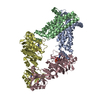 3tuzC 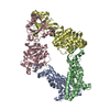 3dhwS S: Starting model for refinement C: citing same article ( |
|---|---|
| Similar structure data |


-Links
 PDBj
PDBj

- Assembly
| Deposited unit | 
| ||||||||
|---|---|---|---|---|---|---|---|---|---|
| 1 | 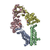
| ||||||||
| 2 | 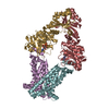
| ||||||||
| Unit cell |
|
-Components
| #1: Protein | Mass: 23269.947 Da / Num. of mol.: 4 Source method: isolated from a genetically manipulated source Source: (gene. exp.) Escherichia coli (E. coli) / Strain: K12 / Gene: b0198, JW0194, metI, yaeE / Production host: Escherichia coli (E. coli) / Strain (production host): BL21* / References: UniProt: P31547#2: Protein | Mass: 40610.191 Da / Num. of mol.: 4 / Mutation: E166Q Source method: isolated from a genetically manipulated source Source: (gene. exp.) Escherichia coli (E. coli) / Strain: K12 / Gene: abc, b0199, JW0195, metN / Production host: Escherichia coli (E. coli) / Strain (production host): BL21*References: UniProt: P30750, Hydrolases; Acting on acid anhydrides; Acting on acid anhydrides to catalyse transmembrane movement of substances#3: Chemical | ChemComp-ADP / Adenosine diphosphate  Mass: 427.201 Da / Num. of mol.: 4 / Source method: obtained synthetically / Formula: C10H15N5O10P2 / Comment: ADP, energy-carrying molecule*YM Mass: 427.201 Da / Num. of mol.: 4 / Source method: obtained synthetically / Formula: C10H15N5O10P2 / Comment: ADP, energy-carrying molecule*YM#4: Water | ChemComp-HOH / | Water Mass: 18.015 Da / Num. of mol.: 146 / Source method: isolated from a natural source / Formula: H2O Mass: 18.015 Da / Num. of mol.: 146 / Source method: isolated from a natural source / Formula: H2O |
|---|
-Experimental details
-Experiment
| Experiment | Method: X-RAY DIFFRACTION / Number of used crystals: 1 |
|---|
- Sample preparation
| Crystal | Density Matthews: 3.42 Å3/Da / Density % sol: 64.07 % |
|---|---|
| Crystal grow | Temperature: 293 K / Method: vapor diffusion, sitting drop / pH: 7.5 Details: 25% v/v polyethylene glycol 400, 0.1 M tris, pH 7.5, VAPOR DIFFUSION, SITTING DROP, temperature 293K |
-Data collection
| Diffraction | Mean temperature: 100 K | |||||||||||||||||||||||||||||||||||||||||||||||||||||||||||||||||||||||||||||
|---|---|---|---|---|---|---|---|---|---|---|---|---|---|---|---|---|---|---|---|---|---|---|---|---|---|---|---|---|---|---|---|---|---|---|---|---|---|---|---|---|---|---|---|---|---|---|---|---|---|---|---|---|---|---|---|---|---|---|---|---|---|---|---|---|---|---|---|---|---|---|---|---|---|---|---|---|---|---|
| Diffraction source | Source: SYNCHROTRON / Site: SSRL  / Beamline: BL12-2 / Wavelength: 1 Å / Beamline: BL12-2 / Wavelength: 1 Å | |||||||||||||||||||||||||||||||||||||||||||||||||||||||||||||||||||||||||||||
| Detector | Type: MARMOSAIC 325 mm CCD / Detector: CCD / Date: Aug 2, 2009 | |||||||||||||||||||||||||||||||||||||||||||||||||||||||||||||||||||||||||||||
| Radiation | Monochromator: Liquid nitrogen-cooled double crystal. / Protocol: SINGLE WAVELENGTH / Monochromatic (M) / Laue (L): M / Scattering type: x-ray | |||||||||||||||||||||||||||||||||||||||||||||||||||||||||||||||||||||||||||||
| Radiation wavelength | Wavelength: 1 Å / Relative weight: 1 | |||||||||||||||||||||||||||||||||||||||||||||||||||||||||||||||||||||||||||||
| Reflection | Redundancy: 3.3 % / Av σ(I) over netI: 12.4 / Number: 303926 / Rsym value: 0.041 / D res high: 2.7 Å / D res low: 29.608 Å / Num. obs: 91606 / % possible obs: 97.3 | |||||||||||||||||||||||||||||||||||||||||||||||||||||||||||||||||||||||||||||
| Diffraction reflection shell |
| |||||||||||||||||||||||||||||||||||||||||||||||||||||||||||||||||||||||||||||
| Reflection | Resolution: 2.7→29.608 Å / Num. all: 91606 / Num. obs: 91606 / % possible obs: 97.3 % / Observed criterion σ(F): -3 / Observed criterion σ(I): 0 / Redundancy: 3.3 % / Rsym value: 0.041 / Net I/σ(I): 12.4 | |||||||||||||||||||||||||||||||||||||||||||||||||||||||||||||||||||||||||||||
| Reflection shell |
|
- Processing
| Software |
| |||||||||||||||||||||||||||||||||||||||||||||||||||||||||||||||||||||||||||||||||||||||||||||||||||||||||||||||||||||||||||||||||||||||||||||||||||||||||||||||||||||||||||||||||||||||||||||
|---|---|---|---|---|---|---|---|---|---|---|---|---|---|---|---|---|---|---|---|---|---|---|---|---|---|---|---|---|---|---|---|---|---|---|---|---|---|---|---|---|---|---|---|---|---|---|---|---|---|---|---|---|---|---|---|---|---|---|---|---|---|---|---|---|---|---|---|---|---|---|---|---|---|---|---|---|---|---|---|---|---|---|---|---|---|---|---|---|---|---|---|---|---|---|---|---|---|---|---|---|---|---|---|---|---|---|---|---|---|---|---|---|---|---|---|---|---|---|---|---|---|---|---|---|---|---|---|---|---|---|---|---|---|---|---|---|---|---|---|---|---|---|---|---|---|---|---|---|---|---|---|---|---|---|---|---|---|---|---|---|---|---|---|---|---|---|---|---|---|---|---|---|---|---|---|---|---|---|---|---|---|---|---|---|---|---|---|---|---|---|
| Refinement | Method to determine structure: MOLECULAR REPLACEMENT Starting model: PDB ENTRY 3DHW Resolution: 2.9→29.609 Å / Occupancy max: 1 / Occupancy min: 0.89 / SU ML: 1.09 / σ(F): 0 / Phase error: 34.29 / Stereochemistry target values: ML
| |||||||||||||||||||||||||||||||||||||||||||||||||||||||||||||||||||||||||||||||||||||||||||||||||||||||||||||||||||||||||||||||||||||||||||||||||||||||||||||||||||||||||||||||||||||||||||||
| Solvent computation | Shrinkage radii: 0.83 Å / VDW probe radii: 1.1 Å / Solvent model: FLAT BULK SOLVENT MODEL / Bsol: 32.985 Å2 / ksol: 0.275 e/Å3 | |||||||||||||||||||||||||||||||||||||||||||||||||||||||||||||||||||||||||||||||||||||||||||||||||||||||||||||||||||||||||||||||||||||||||||||||||||||||||||||||||||||||||||||||||||||||||||||
| Displacement parameters |
| |||||||||||||||||||||||||||||||||||||||||||||||||||||||||||||||||||||||||||||||||||||||||||||||||||||||||||||||||||||||||||||||||||||||||||||||||||||||||||||||||||||||||||||||||||||||||||||
| Refinement step | Cycle: LAST / Resolution: 2.9→29.609 Å
| |||||||||||||||||||||||||||||||||||||||||||||||||||||||||||||||||||||||||||||||||||||||||||||||||||||||||||||||||||||||||||||||||||||||||||||||||||||||||||||||||||||||||||||||||||||||||||||
| Refine LS restraints |
| |||||||||||||||||||||||||||||||||||||||||||||||||||||||||||||||||||||||||||||||||||||||||||||||||||||||||||||||||||||||||||||||||||||||||||||||||||||||||||||||||||||||||||||||||||||||||||||
| LS refinement shell |
|
