 Movie
Movie Controller

[English] 日本語
 Yorodumi
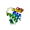
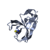
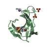
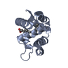
- PDB-3tsv: crystal structure of the third PDZ domain of the human ZO-1 MAGUK... -
+ Open data

- Basic information
| Entry | Database: PDB / ID: 3tsv | ||||||
|---|---|---|---|---|---|---|---|
| Title | crystal structure of the third PDZ domain of the human ZO-1 MAGUK protein | ||||||
 Components Components | Tight junction protein ZO-1 | ||||||
 Keywords Keywords | CELL ADHESION / PDZ / Scaffolding / JAM / Tight junction | ||||||
| Function / homology |  Function and homology information Function and homology informationpositive regulation of blood-brain barrier permeability / adherens junction maintenance / positive regulation of cell-cell adhesion mediated by cadherin / RUNX1 regulates expression of components of tight junctions / SARS-CoV-2 targets PDZ proteins in cell-cell junction / establishment of endothelial intestinal barrier / protein localization to cell-cell junction / regulation of cell junction assembly / Regulation of gap junction activity / protein localization to bicellular tight junction ...positive regulation of blood-brain barrier permeability / adherens junction maintenance / positive regulation of cell-cell adhesion mediated by cadherin / RUNX1 regulates expression of components of tight junctions / SARS-CoV-2 targets PDZ proteins in cell-cell junction / establishment of endothelial intestinal barrier / protein localization to cell-cell junction / regulation of cell junction assembly / Regulation of gap junction activity / protein localization to bicellular tight junction / protein localization to adherens junction / gap junction / cell-cell junction organization / actomyosin structure organization / Apoptotic cleavage of cell adhesion proteins / podosome / Signaling by Hippo / tight junction / regulation of bicellular tight junction assembly / cell-cell junction assembly / negative regulation of stress fiber assembly / apical junction complex / maintenance of blood-brain barrier / positive regulation of sprouting angiogenesis / regulation of cytoskeleton organization / bicellular tight junction / cell adhesion molecule binding / cell projection / adherens junction / cell-cell adhesion / apical part of cell / cell junction / actin cytoskeleton organization / basolateral plasma membrane / calmodulin binding / positive regulation of cell migration / cadherin binding / positive regulation of cell population proliferation / negative regulation of apoptotic process / protein-containing complex / plasma membrane / cytosol / cytoplasmSimilarity search - Function | ||||||
| Biological species |  Homo sapiens (human) Homo sapiens (human) | ||||||
| Method | X-RAY DIFFRACTION / SYNCHROTRON / MOLECULAR REPLACEMENT / Resolution: 1.989 Å | ||||||
 Authors Authors | Nomme, J. / Lavie, A. | ||||||
 Citation Citation | Journal: J.Biol.Chem. / Year: 2011 Title: The Src Homology 3 Domain Is Required for Junctional Adhesion Molecule Binding to the Third PDZ Domain of the Scaffolding Protein ZO-1. Authors: Nomme, J. / Fanning, A.S. / Caffrey, M. / Lye, M.F. / Anderson, J.M. / Lavie, A. | ||||||
| History |
|
- Structure visualization
| Structure viewer | Molecule: MolmilJmol/JSmol |
|---|
- Downloads & links
-Download
| PDBx/mmCIF format | 3tsv.cif.gz | 31 KB | Display | PDBx/mmCIF format |
|---|---|---|---|---|
| PDB format | pdb3tsv.ent.gz | 20 KB | Display | PDB format |
| PDBx/mmJSON format | 3tsv.json.gz | Tree view | PDBx/mmJSON format | |
| Others |  Other downloads Other downloads |
-Validation report
| Arichive directory | https://data.pdbj.org/pub/pdb/validation_reports/ts/3tsvftp://data.pdbj.org/pub/pdb/validation_reports/ts/3tsv | HTTPS FTP |
|---|
-Related structure data
| Related structure data |  3tswC  3tszC 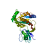 3lh5S C: citing same article ( S: Starting model for refinement |
|---|---|
| Similar structure data |


-Links
 PDBj
PDBj


- Assembly
| Deposited unit | 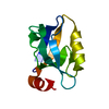
| ||||||||
|---|---|---|---|---|---|---|---|---|---|
| 1 |
| ||||||||
| Unit cell |
|
-Components
| #1: Protein | / Tight junction protein 1 / Zona occludens protein 1 / Zonula occludens protein 1 Mass: 13710.578 Da / Num. of mol.: 1 / Fragment: PDZ3 domain (UNP Residues 417-516) Source method: isolated from a genetically manipulated source Source: (gene. exp.) Homo sapiens (human) / Gene: TJP1, ZO1 / Plasmid: pET14b / Production host:  Escherichia coli (E. coli) / Strain (production host): BL21(C41) / References: UniProt: Q07157 Escherichia coli (E. coli) / Strain (production host): BL21(C41) / References: UniProt: Q07157 |
|---|---|
| #2: Water | ChemComp-HOH / Water Mass: 18.015 Da / Num. of mol.: 32 / Source method: isolated from a natural source / Formula: H2O Mass: 18.015 Da / Num. of mol.: 32 / Source method: isolated from a natural source / Formula: H2O |
-Experimental details
-Experiment
| Experiment | Method: X-RAY DIFFRACTION / Number of used crystals: 1 |
|---|
- Sample preparation
| Crystal | Density Matthews: 1.69 Å3/Da / Density % sol: 27.3 % |
|---|---|
| Crystal grow | Temperature: 293.15 K / Method: vapor diffusion, sitting drop / pH: 5 Details: 25% PEG 1500, 0.1M MMT, pH 5.0, VAPOR DIFFUSION, SITTING DROP, temperature 293.15K |
-Data collection
| Diffraction | Mean temperature: 100 K |
|---|---|
| Diffraction source | Source: SYNCHROTRON / Site: APS  / Beamline: 22-BM / Wavelength: 1 Å / Beamline: 22-BM / Wavelength: 1 Å |
| Detector | Type: MARMOSAIC 225 mm CCD / Detector: CCD / Date: Mar 18, 2011 |
| Radiation | Protocol: SINGLE WAVELENGTH / Monochromatic (M) / Laue (L): M / Scattering type: x-ray |
| Radiation wavelength | Wavelength: 1 Å / Relative weight: 1 |
| Reflection | Resolution: 1.989→35.98 Å / Num. all: 6649 / Num. obs: 6649 / % possible obs: 97.8 % / Observed criterion σ(F): 0 / Observed criterion σ(I): 0 / Redundancy: 3.2 % / Rmerge(I) obs: 0.05 / Net I/σ(I): 15.1 |
| Reflection shell | Resolution: 1.989→2.11 Å / Redundancy: 3.2 % / Rmerge(I) obs: 0.61 / Mean I/σ(I) obs: 2.1 / Num. unique all: 995 / % possible all: 93.3 |
- Processing
| Software |
| |||||||||||||||||||||||||||||||||||||||||||||||||||||||||||||||||
|---|---|---|---|---|---|---|---|---|---|---|---|---|---|---|---|---|---|---|---|---|---|---|---|---|---|---|---|---|---|---|---|---|---|---|---|---|---|---|---|---|---|---|---|---|---|---|---|---|---|---|---|---|---|---|---|---|---|---|---|---|---|---|---|---|---|---|
| Refinement | Method to determine structure: MOLECULAR REPLACEMENT Starting model: PDB ENTRY 3LH5 Resolution: 1.989→35.98 Å / Cor.coef. Fo:Fc: 0.926 / Cor.coef. Fo:Fc free: 0.917 / SU B: 8.19 / SU ML: 0.226 / Cross valid method: THROUGHOUT / σ(F): 0 / ESU R: 0.281 / ESU R Free: 0.232 / Stereochemistry target values: MAXIMUM LIKELIHOOD / Details: HYDROGENS HAVE BEEN ADDED IN THE RIDING POSITIONS
| |||||||||||||||||||||||||||||||||||||||||||||||||||||||||||||||||
| Solvent computation | Ion probe radii: 0.8 Å / Shrinkage radii: 0.8 Å / VDW probe radii: 1.4 Å / Solvent model: MASK | |||||||||||||||||||||||||||||||||||||||||||||||||||||||||||||||||
| Displacement parameters | Biso mean: 36.873 Å2
| |||||||||||||||||||||||||||||||||||||||||||||||||||||||||||||||||
| Refinement step | Cycle: LAST / Resolution: 1.989→35.98 Å
| |||||||||||||||||||||||||||||||||||||||||||||||||||||||||||||||||
| Refine LS restraints |
| |||||||||||||||||||||||||||||||||||||||||||||||||||||||||||||||||
| LS refinement shell | Resolution: 1.989→2.04 Å / Total num. of bins used: 20
|
