 Movie
Movie Controller

[English] 日本語
 Yorodumi
- PDB-3tny: Structure of YfiY from Bacillus cereus bound to the siderophore i... -
+ Open data

- Basic information
| Entry | Database: PDB / ID: 3tny | ||||||
|---|---|---|---|---|---|---|---|
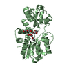
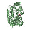
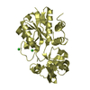
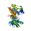
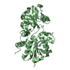
| Title | Structure of YfiY from Bacillus cereus bound to the siderophore iron (III) schizokinen | ||||||
 Components Components | YfiY (ABC transport system substrate-binding protein) | ||||||
 Keywords Keywords | METAL TRANSPORT / schizokinen /  bacillus cereus / nutrient binding protein / ABC transporter bacillus cereus / nutrient binding protein / ABC transporter | ||||||
| Function / homology | Nitrogenase molybdenum iron protein domain / Rossmann fold / 3-Layer(aba) Sandwich / Alpha Beta / Chem-SKZ / :  Function and homology information Function and homology information | ||||||
| Biological species |  Bacillus cereus (bacteria) Bacillus cereus (bacteria) | ||||||
| Method | X-RAY DIFFRACTION / SYNCHROTRON / MOLECULAR REPLACEMENT / molecular replacement / Resolution: 1.55 Å | ||||||
 Authors Authors | Clifton, M.C. | ||||||
 Citation Citation | Journal: To be Published Title: Parsing the functional specificity of Siderocalin / Lipocalin 2 / NGAL for siderophores and related small-molecule ligands Authors: Clifton, M.C. / Rupert, P.B. / Hoette, T.M. / Raymond, K.N. / Abergel, R.J. / Strong, R.K. | ||||||
| History |
|
- Structure visualization
| Structure viewer | Molecule: MolmilJmol/JSmol |
|---|
- Downloads & links
-Download
| PDBx/mmCIF format | 3tny.cif.gz | 77.5 KB | Display | PDBx/mmCIF format |
|---|---|---|---|---|
| PDB format | pdb3tny.ent.gz | 56.2 KB | Display | PDB format |
| PDBx/mmJSON format | 3tny.json.gz | Tree view | PDBx/mmJSON format | |
| Others |  Other downloads Other downloads |
-Validation report
| Arichive directory | https://data.pdbj.org/pub/pdb/validation_reports/tn/3tnyftp://data.pdbj.org/pub/pdb/validation_reports/tn/3tny | HTTPS FTP |
|---|
-Related structure data
| Related structure data | 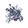 3cmpC  3hwdC 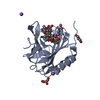 3hweC 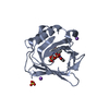 3hwfC 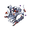 3hwgC  3i0aC 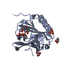 3k3lC  3tf6C  3tzsC C: citing same article ( |
|---|---|
| Similar structure data |


-Links
 PDBj
PDBj- Assembly
| Deposited unit | 
| ||||||||
|---|---|---|---|---|---|---|---|---|---|
| 1 |
| ||||||||
| Unit cell |
|
-Components
| #1: Protein | Mass: 34460.273 Da / Num. of mol.: 1 Source method: isolated from a genetically manipulated source Source: (gene. exp.) Bacillus cereus (bacteria) / Gene: yfiY / Plasmid: pET101 / Production host: Escherichia coli (E. coli) / References: UniProt: C2Y5Z3 |
|---|---|
| #2: Chemical | ChemComp-SKZ / [  Mass: 470.212 Da / Num. of mol.: 1 / Source method: obtained synthetically / Formula: C16H22FeN4O9 Mass: 470.212 Da / Num. of mol.: 1 / Source method: obtained synthetically / Formula: C16H22FeN4O9 |
| #3: Water | ChemComp-HOH / Water Mass: 18.015 Da / Num. of mol.: 374 / Source method: isolated from a natural source / Formula: H2O Mass: 18.015 Da / Num. of mol.: 374 / Source method: isolated from a natural source / Formula: H2O |
-Experimental details
-Experiment
| Experiment | Method: X-RAY DIFFRACTION / Number of used crystals: 1 |
|---|
- Sample preparation
| Crystal | Density Matthews: 2.69 Å3/Da / Density % sol: 54.2 % |
|---|---|
| Crystal grow | Temperature: 312 K / Method: vapor diffusion, hanging drop / pH: 7 Details: Protein 10-20 mg/ml, 0.1M HEPES 7.0, 30% Jeffamine ED-2001. Direct cryoprotection. , VAPOR DIFFUSION, HANGING DROP, temperature 312K |
-Data collection
| Diffraction | Mean temperature: 100 K |
|---|---|
| Diffraction source | Source: SYNCHROTRON / Site: ALS  / Beamline: 5.0.2 / Wavelength: 0.97 Å / Beamline: 5.0.2 / Wavelength: 0.97 Å |
| Detector | Type: ADSC QUANTUM 315 / Detector: CCD / Date: May 1, 2010 |
| Radiation | Monochromator: Liquid Nitrogen cooled Dual Crystal / Protocol: SINGLE WAVELENGTH / Monochromatic (M) / Laue (L): M / Scattering type: x-ray |
| Radiation wavelength | Wavelength: 0.97 Å / Relative weight: 1 |
| Reflection | Resolution: 1.55→50 Å / Num. obs: 46458 / % possible obs: 99.5 % / Redundancy: 7.3 % / Rmerge(I) obs: 0.118 / Χ2: 0.949 / Net I/σ(I): 9.2 |
| Reflection shell | Resolution: 1.55→1.61 Å / Χ2: 1.001 |
-Phasing
| Phasing | Method: molecular replacement |
|---|
- Processing
| Software |
| |||||||||||||||||||||||||||||||||||||||||||||||||||||||||||||||||||||||||||||||||||||
|---|---|---|---|---|---|---|---|---|---|---|---|---|---|---|---|---|---|---|---|---|---|---|---|---|---|---|---|---|---|---|---|---|---|---|---|---|---|---|---|---|---|---|---|---|---|---|---|---|---|---|---|---|---|---|---|---|---|---|---|---|---|---|---|---|---|---|---|---|---|---|---|---|---|---|---|---|---|---|---|---|---|---|---|---|---|---|
| Refinement | Method to determine structure: MOLECULAR REPLACEMENT / Resolution: 1.55→50 Å / Cor.coef. Fo:Fc: 0.968 / Cor.coef. Fo:Fc free: 0.961 / WRfactor Rfree: 0.2085 / WRfactor Rwork: 0.1868 / Occupancy max: 1 / Occupancy min: 0.3 / FOM work R set: 0.8909 / SU B: 1.21 / SU ML: 0.044 / SU R Cruickshank DPI: 0.0758 / SU Rfree: 0.0727 / Cross valid method: THROUGHOUT / σ(F): 0 / ESU R Free: 0.073 / Stereochemistry target values: MAXIMUM LIKELIHOOD Details: HYDROGENS HAVE BEEN ADDED IN THE RIDING POSITIONS U VALUES : REFINED INDIVIDUALLY
| |||||||||||||||||||||||||||||||||||||||||||||||||||||||||||||||||||||||||||||||||||||
| Solvent computation | Ion probe radii: 0.8 Å / Shrinkage radii: 0.8 Å / VDW probe radii: 1.2 Å / Solvent model: MASK | |||||||||||||||||||||||||||||||||||||||||||||||||||||||||||||||||||||||||||||||||||||
| Displacement parameters | Biso max: 56.47 Å2 / Biso mean: 21.1498 Å2 / Biso min: 12.36 Å2
| |||||||||||||||||||||||||||||||||||||||||||||||||||||||||||||||||||||||||||||||||||||
| Refinement step | Cycle: LAST / Resolution: 1.55→50 Å
| |||||||||||||||||||||||||||||||||||||||||||||||||||||||||||||||||||||||||||||||||||||
| Refine LS restraints |
| |||||||||||||||||||||||||||||||||||||||||||||||||||||||||||||||||||||||||||||||||||||
| LS refinement shell | Resolution: 1.55→1.59 Å / Total num. of bins used: 20
|
